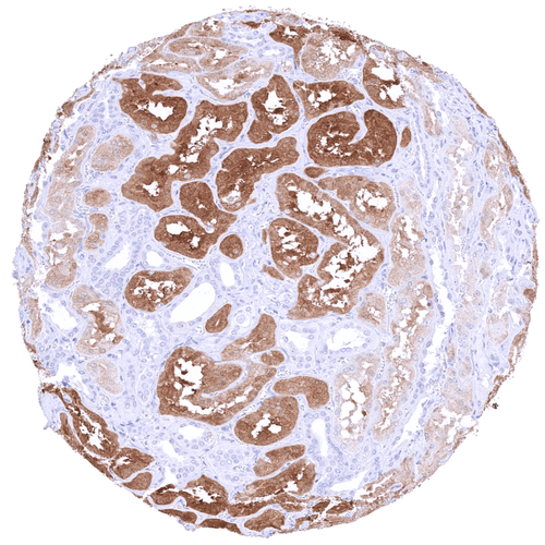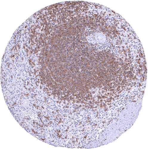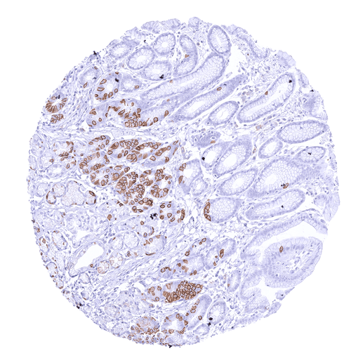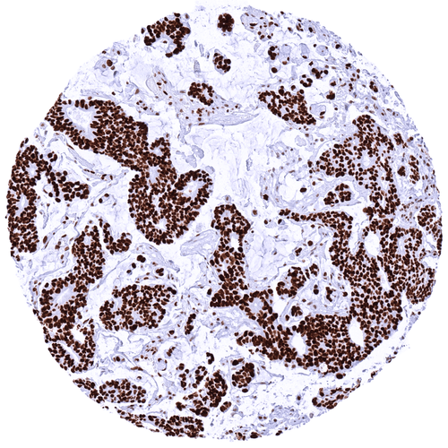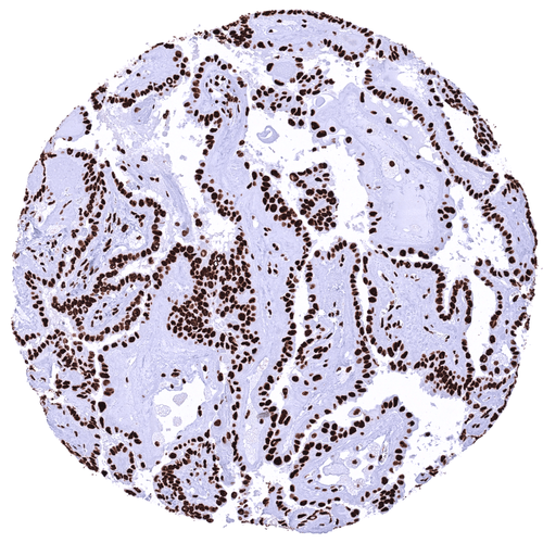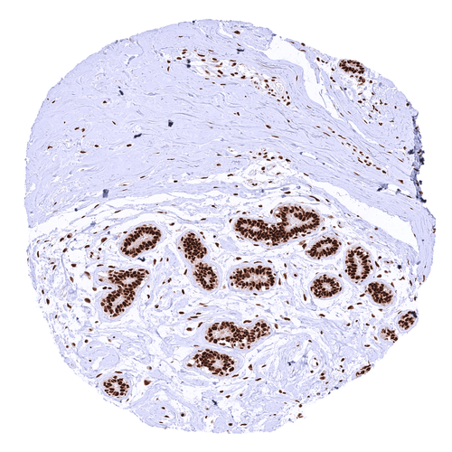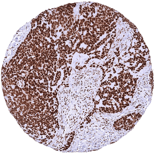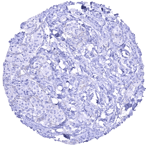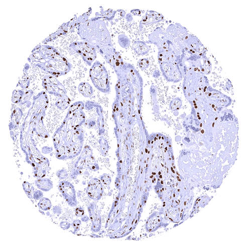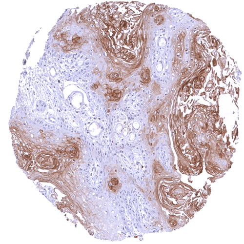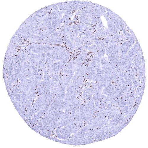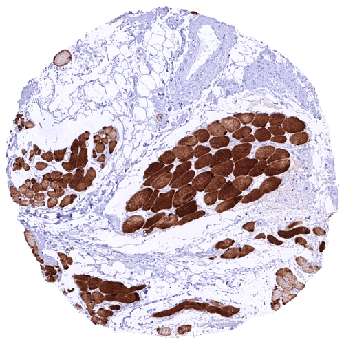

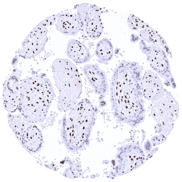
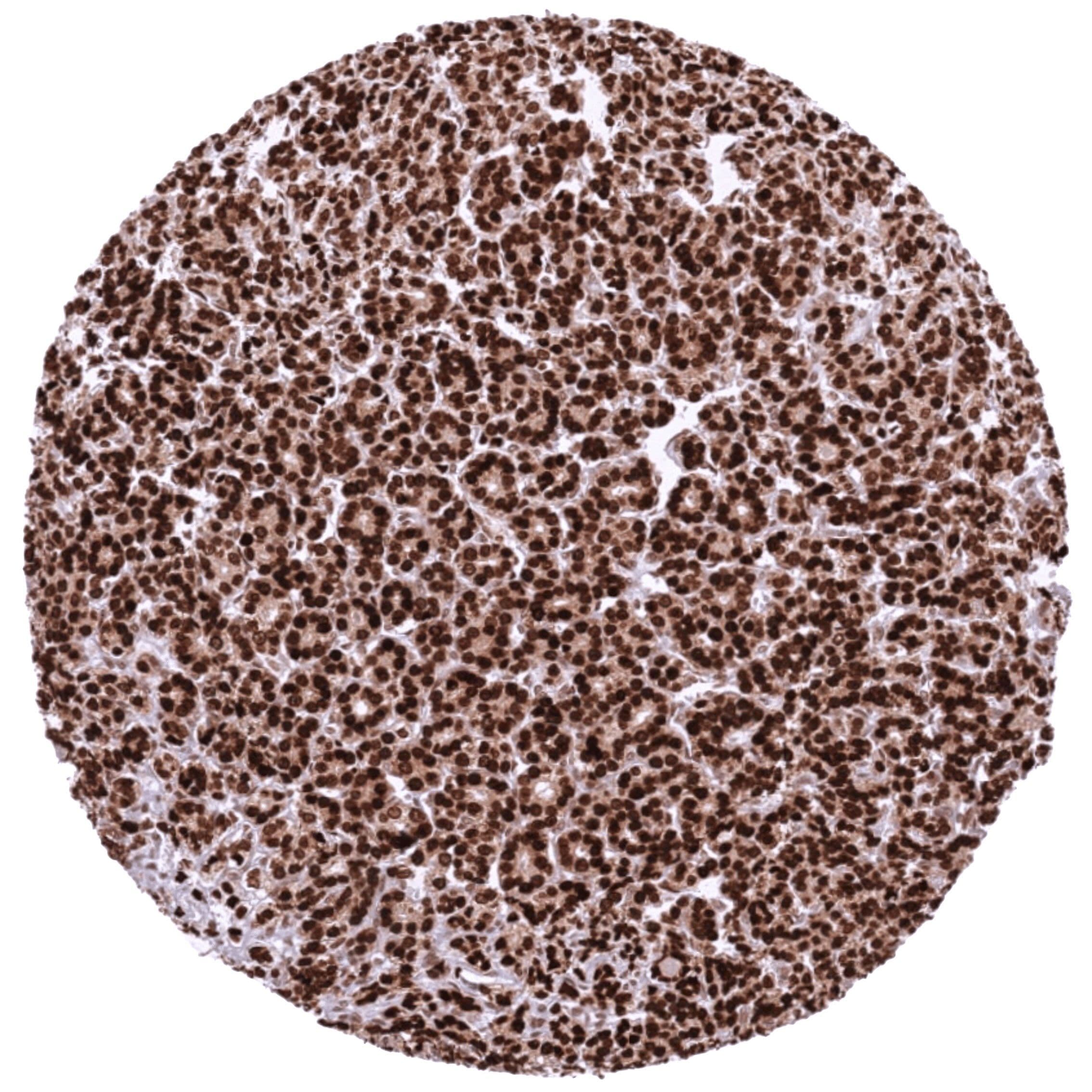
Follicular carcinoma of the thyroid showing a strong nuclear HMGA2 staining of all tumor cells
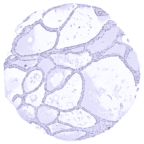
Staining Pattern in Normal Tissues
Manual protocol
Freshly cut sections should be used (less than 10 days between cutting and staining). Heat-induced antigen retrieval for 5 minutes in an autoclave at 121°C in pH 7,8 Target Retrieval Solution buffer. Apply HMV314 at a dilution of 1:150 at 37°C for 60 minutes. Visualization of bound antibody by the EnVision Kit (Dako, Agilent) according to the manufacturer’s directions.
| Brain | Cerebrum, grey | Negative | |
| Cerebrum, white | Negative | ||
| Cerebellum, cortex | Negative | ||
| Cerebellum, white | Negative | ||
| Ganglion | Negative | ||
| Ependyma | Negative | ||
| Eye, retina | Negative | ||
| Endocrine Tissues | Thyroid | Most follicular cells show a faint to weak nuclear HMGA2 staining. | |
| Parathyroid gland | Some epithelial cell groups show a moderate to strong nuclear HMGA2 staining | ||
| Adrenal gland | Negative | ||
| Pituitary gland, anterior lobe | Weak to moderate nuclear HMGA2 staining of pituicytes. Adenohypophysis is HMGA2 negative. | ||
| Pituitary gland, posterior lobe | Negative | ||
| Respiratory system | Lung bronchi | Negative | |
| Lung, bronchial glands | Negative | ||
| Nose, paranasal sinus | Negative | ||
| Lung, parenchyma | Negative | ||
| Proximal digestive tract | Lip | Negative | |
| Oral cavity | Negative | ||
| Tonsil, surface | Negative | ||
| Esophagus, mucosa | Negative | ||
| Lip, small salivary gland | Negative | ||
| Sublingual gland | Negative | ||
| Parotid gland | Negative | ||
| Submandibullary gland | Negative | ||
| Gastronintestinal tract | Stomach, antrum | Negative | |
| Stomach, fundus and corpus | Negative | ||
| Small intestine, duodenum | Negative | ||
| Duodenum, Brunner gland | Weak nuclear HMGA2 staining of duodenal crypt cells. Moderate HMGA2 positivity of Brunner glands. | ||
| Small intestine, ileum | Negative | ||
| Appendix | Negative | ||
| Colon descendens | Negative | ||
| Rectum | Negative | ||
| Anal canal, transition epithelium | Negative | ||
| Liver, Gallbladder, Pancreas | Liver | Weak nuclear HMGA2 staining of a fraction of bile duct epithelial cells. Hepatocytes are negative. | |
| Gallbladder | Negative | ||
| Pancreas | Weak nuclear HMGA2 staining of a fraction of acinar and ductal (small ducts) cells. | ||
| Kidney, urinary bladder | Kidney, cortex | Moderate nuclear HMGA2 staining of tubular cells in areas of tissue damage. Faint nuclear HMGA2 staining of a fraction of collecting duct cells. | |
| Kidney, medulla | Negative | ||
| Urinary bladder, urothelium | Few urothelial cells show a weak nuclear HMGA2 positivity. | ||
| Kidney pelvis, mucosa | Negative | ||
| Male tissues | Prostate | Negative | |
| Seminal vesicle | Strong nuclear HMGA2 staining of a large fraction of acinar glandular cells. | ||
| Epididymis caput | Weak nuclear HMGA2 staining of a fraction of epithelial cells in the cauda while cells in the corpus are HMGA2 negative. | ||
| Epididymis cauda | Negative | ||
| Testis | Weak to moderate nuclear HMGA2 staining of a spermatocytes and of spermatozoa. | ||
| Female Tissues | Breast, glands | Negative | |
| Ectocervix | Negative | ||
| Endocervix | Strong nuclear HMGA2 staining of epithelial cells. | ||
| Endometrium, proliferation | Negative | ||
| Endometrium, secretion | Weak to moderate nuclear HMGA2 staining of a subset of epithelial cells. | ||
| Uterus, myometrium | Negative | ||
| Fallopian tube | Strong nuclear HMGA2 staining of most epithelial cells. | ||
| Ovary, stroma | Negative | ||
| Ovary, follicular cyst | Negative | ||
| Ovary, corpus luteum | Negative | ||
| Amnion | Negative | ||
| Chorion | Negative | ||
| Amnion/Chorion | Strong nuclear HMGA2 positivity of amnion cells. | ||
| Placenta, early, decidua | Strong nuclear HMGA2 staining of stroma cells. Trophoblast cells remain HMGA2 negative. | ||
| Placenta, first trimenon | Negative | ||
| Placenta, mature | Some nuclear HMGA2 staining of trophoblast cells in some samples. Stroma cells are HMGA2 negative. | ||
| Muscle, connective & soft tissue | Aorta, intima | Negative | |
| Skeletal muscle | Weak HMGA2 staining of some myocytes | ||
| Aorta, media | Negative | ||
| Skeletal muscle, tongue | Negative | ||
| Heart, left ventricle | Negative | ||
| Kidney pelvis, muscular wall | Negative | ||
| Urinary bladder, muscular wall | Negative | ||
| Esophagus, muscular wall | Negative | ||
| Stomach, muscular wall | Negative | ||
| Ileum, muscular wall | Negative | ||
| Appendix, muscular wall | Negative | ||
| Colon descendens, muscular wall | Negative | ||
| Penis, glans, corpus spongiosum | Negative | ||
| Fat, white | Negative | ||
| Skin | Skin, surface | Negative | |
| Skin (hairs, sebaceous glands) | Negative | ||
| Anal canal, skin | Negative | ||
| Scrotum | Negative | ||
| Bone Marrow & lymphoid tissues | Bone marrow | Moderate nuclear HMGA2 staining of a fraction of cells. | |
| Thymus | Negative | ||
| Spleen | Negative | ||
| Lymph node | Negative | ||
| Tonsil, deep | Negative |
Details
More product details
More product details
Biology behind
The high mobility group protein 2 (HMGA2) is a protein of less than 12 kDa, that consists of 108 amino acids and which is coded by the HMGA2 gene located at 12q13-15. The first three (of five) exons encode the AT-binding domain site, which can bind to AT-rich binding sites in the DNA minor groove. These bindings affect the conformation of the DNA and modify transcription by enhancing or suppressing the activities of numerous genes. As such, HMGA2 is an essential component of the enhanceosome. Instead of direct regulation, HMGA2 alters the architecture of DNA and supports the assembly of protein complexes that do regulate the transcription of genes. HMGA2 is preferentially expressed during organogenesis and – with few exceptions – it is hardly expressed in adult tissues. HMGA2 appears to be essential for cell growth regulation. A knock-out of the HMGA2 counterpart in mice results in diet-induced obesity. Specific HMGA2 mutations can lead to unusually small size in mice. Genome-wide association studies have found a relationship between height of humans and HMGA2-linked SNPs. Data suggesting a role of HMGA2 in cancer are rapidly accumulating. Aberrant expression of HMGA2 in adult tissues is commonly associated with both benign and malignant tumor formation. HMGA2 is often re-expressed in human tumors, where it promotes tumorigenesis by multiple mechanisms. HMGA2 was found to increase cancer cell proliferation, inhibit apoptosis, impact several DNA repair mechanisms, endorse epithelial-mesenchymal transition, support a cancer stem cell phenotype, and to foster cancer cell resistance to chemotherapeutic agents. Causes for HMGA2 re-expression in neoplastic tissues include gene fusion, amplification, regulation by specific miRNAs, and other mechanisms.
Protocol Recommendations
Protocol Recommendations
Potential Research Applications
Potential Research Applications
Evidence For Specificity In I H C
Evidence For Specificity In I H C