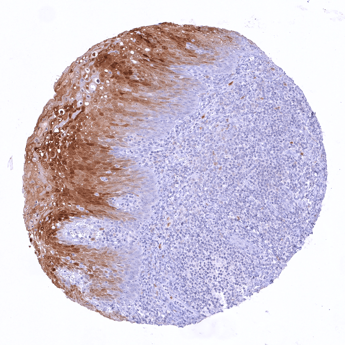
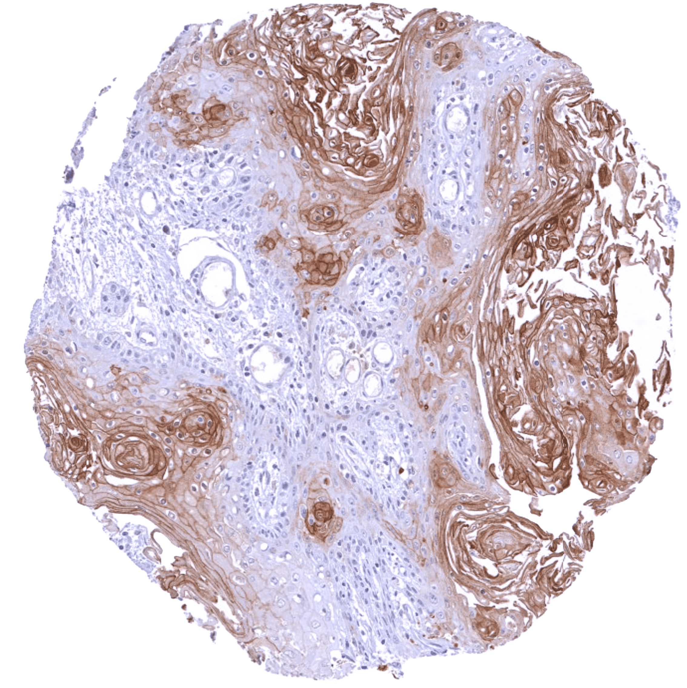
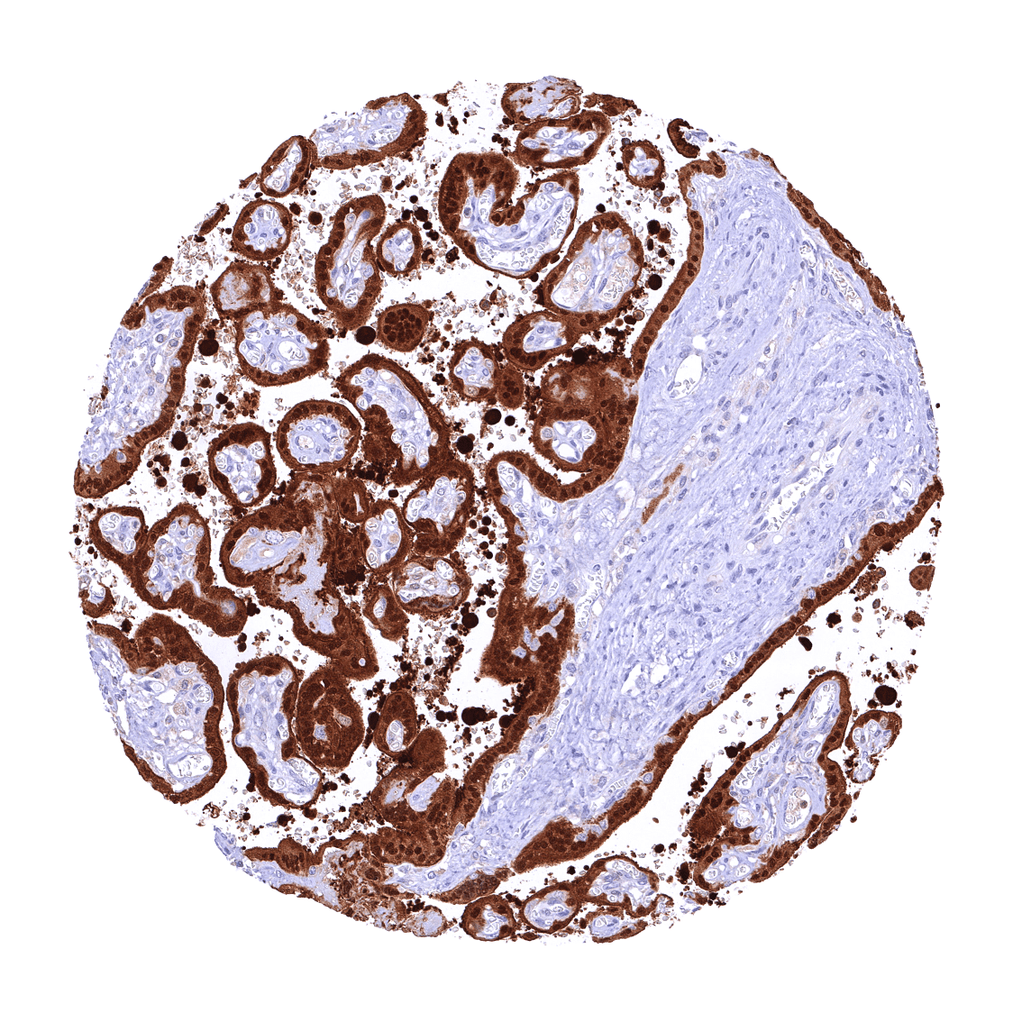
Staining Pattern in Normal Tissues
Manual protocol
Freshly cut sections should be used (less than 10 days between cutting and staining). Heat-induced antigen retrieval for 5 minutes in an autoclave at 121°C in pH 7,8 Target Retrieval Solution buffer. Apply HMV330 at a dilution of 1:200 at 37°C for 60 minutes. Visualization of bound antibody by the EnVision Kit (Dako, Agilent) according to the manufacturer’s directions.
Brain | Cerebrum | Negative | |
Cerebellum | Negative | ||
Endocrine tissues | Thyroid | Negative | |
Parathyroid | Negative | ||
Adrenal gland | Negative | ||
Pituitary gland | Negative | ||
Respiratory system | Respiratory epithelium | Negative | |
Lung | Negative | ||
Gastrointestinal tract | Salivary glands | Moderate to strong cytoplasmic and nuclear PAI2 staining of epithelial cells from excretory ducts (not in all samples). | |
Esophagus | Weak cytoplasmic and nuclear PAI2 staining of top 2/3 cell layers of squamous epithelium (basal and suprabasal layers are PAI2 negative). | ||
Stomach | Negative | ||
Duodenum | Negative | ||
Small intestine | Negative | ||
Appendix | Negative | ||
Colon | Negative | ||
Rectum | Negative | ||
Liver, Gallbladder, Pancreas | Liver | Negative | |
Gallbladder | Negative | ||
Pancreas | Moderate to strong cytoplasmic and nuclear PAI2 staining of a small subset of epithelial cells. | ||
Genitourinary | Kidney | Negative | |
Urothelium | Negative | ||
Male tissues | Prostate | Negative | |
Seminal Vesicles | Negative | ||
Testis | Negative | ||
Epididymis | Negative | ||
Female tissues | Breast | Negative | |
Uterus, myometrium | Negative | ||
Uterus, ectocervix | Weak cytoplasmic and nuclear PAI2 staining of intermediate layers of squamous epithelium (superficial and basal layers are PAI2 negative). | ||
Uterus, endocervix | Negative | ||
Uterus, endometrium | Negative | ||
Fallopian tube | Negative | ||
Ovary | Weak cytoplasmic and nuclear PAI2 staining of few dispersed corpus luteum cells. | ||
Placenta early | Strong cytoplasmic and nuclear PAI2 staining of trophoblast cells. Strong PAI2 staining of decidua cells. | ||
Placenta mature | Strong cytoplasmic and nuclear PAI2 staining of trophoblast cells. Strong PAI2 staining of decidua cells. | ||
Amnion | Strong PAI2 staining of amnion cells | ||
Chorion | Negative | ||
Skin | Epidermis | At least n some samples, there is a moderate to strong predominantly nuclear PAI2 staining of the lower 2/3 of the squamous epithelium (including the basal cell layer). | |
Sebaceous glands | Negative | ||
Muscle, connective & soft tissues | Heart muscle | Negative | |
Skeletal muscle | Negative | ||
Smooth muscle | Negative | ||
Vessel walls | Negative | ||
Fat | Negative | ||
Stroma | Negative | ||
Endothelium | Negative | ||
Bone marrow & lymphoid tissues | Bone marrow | Very few cells show a nuclear and cytoplasmic PAI2 staining. | |
Lymph node | Negative | ||
Spleen | Negative | ||
Thymus | Moderate to strong PAI2 staining of a subset of cells of corpuscles of Hassall‘s. | ||
Tonsil | Strong cytoplasmic and nuclear PAI2 staining of the upper 1/2 to-2/3 of the squamous epithelium. | ||
Remarks |
Details
More product details
Biology behind
Plasminogen activator inhibitor-2 (placental PAI, SerpinB2, PAI-2) is a serine protease inhibitor of the serpin superfamily coded by the SerpinB2 gene on chromosome 18.q22.1. It acts as a coagulation factor that irreversibly inactivates tissue plasminogen activator and urokinase. PAI2 exists as a 60-kDa extracellular glycosylated form and a 43-kDa intracellular form. PAI2 protein is normally not detectable in adult plasma. The protein is produced at large quantities in the placenta which explains that PAI2 is detectable in blood only during pregnancy. PAI2 may thus contribute to the increased rate of thrombosis during pregnancy. PAI2 can bind to multiple intracellular and extracellular proteins. For example, it was suggested that PAI2 may activate p53 and stabilize p21. Macrophage derived PAI2 plays a role in inflammatory responses and infections, potentially in downregulating T cells that secrete IgG2c and interferon type II. Although glycosylated extracellular PAI2 regulates fibrinolysis, it remains unclear whether this is the main or entire role of PAI2. PAI2 is predominantly intracellular but specific intracellular roles for PAI2 have not yet been identified. PAI2 may play a complex role in cancer. Both tumor-promoting and tumor-inhibiting effects have been described.
Protocol Recommendations
Potential Research Applications
Evidence For Antibody Specificity In I H C