
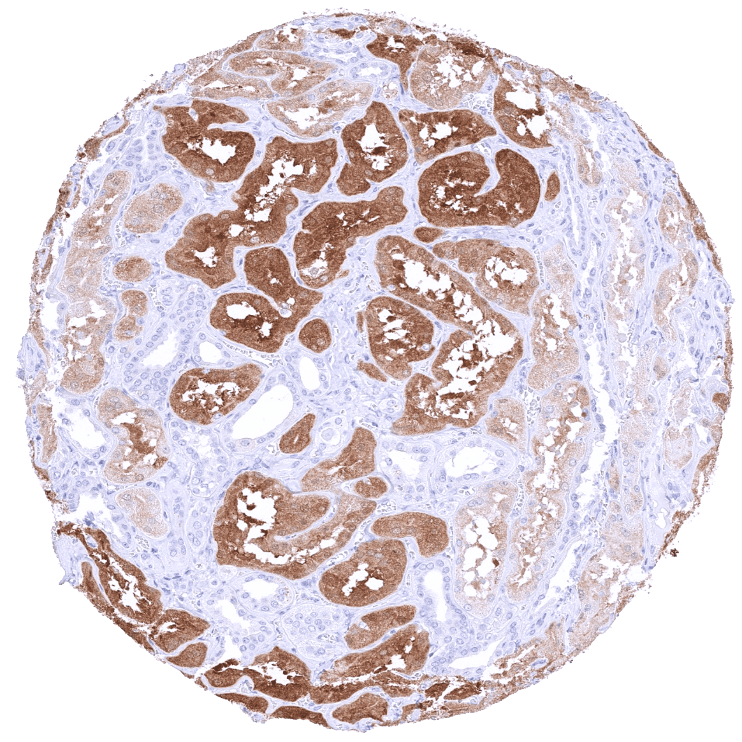
Kidney displaying a predominantly cytoplasmic PSAT1 staining of variable intensity in most tubuli
Staining Pattern in Normal Tissues
Manual protocol
Freshly cut sections should be used (less than 10 days between cutting and staining). Heat-induced antigen retrieval for 5 minutes in an autoclave at 121°C in pH 7,8 Target Retrieval Solution buffer. Apply HMV331 at a dilution of 1:200 at 37°C for 60 minutes. Visualization of bound antibody by the EnVision Kit (Dako, Agilent) according to the manufacturer’s directions.
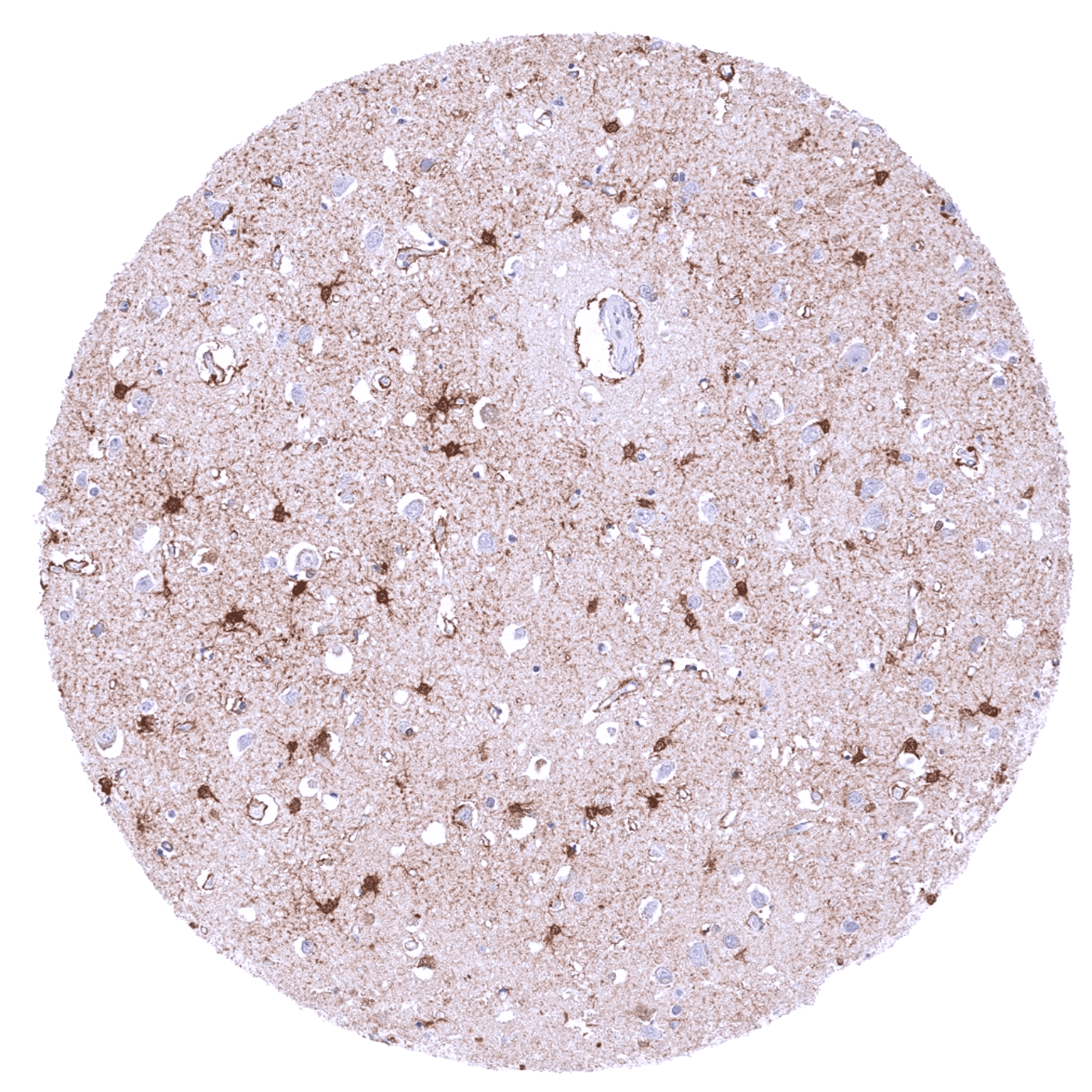
| Brain | Cerebrum, grey | Negative | |
| Cerebrum, white | Negative | ||
| Cerebellum, cortex | Negative | ||
| Cerebellum, white | Negative | ||
| Ganglion | Negative | ||
| Ependyma | Negative | ||
| Eye, retina | Negative | ||
| Endocrine Tissues | Thyroid | Negative | |
| Parathyroid gland | Weak to moderate, predominantly cytoplasmic PSAT1 staining of a small subset of cells (not in all samples). | ||
| Adrenal gland | Weak to moderate, predominantly cytoplasmic PSAT1 staining of a subset of cells. | ||
| Pituitary gland, anterior lobe | Negative | ||
| Pituitary gland, posterior lobe | Negative | ||
| Respiratory system | Lung bronchi | Negative | |
| Lung, bronchial glands | Negative | ||
| Nose, paranasal sinus | Negative | ||
| Lung, parenchyma | Negative | ||
| Proximal digestive tract | Lip | Negative | |
| Oral cavity | Negative | ||
| Tonsil, surface | Negative | ||
| Esophagus, mucosa | Negative | ||
| Lip, small salivary gland | Negative | ||
| Sublingual gland | Negative | ||
| Parotid gland | Negative | ||
| Submandibullary gland | Negative | ||
| Gastronintestinal tract | Stomach, antrum | Negative | |
| Stomach, fundus and corpus | Negative | ||
| Small intestine, duodenum | Negative | ||
| Duodenum, Brunner gland | Negative | ||
| Small intestine, ileum | Negative | ||
| Appendix | Distinct cytoplasmic and nuclear PSAT1 staining of few crypt cells. | ||
| Colon descendens | Negative | ||
| Rectum | Distinct cytoplasmic and nuclear PSAT1 staining of few crypt cells. | ||
| Anal canal, transition epithelium | Negative | ||
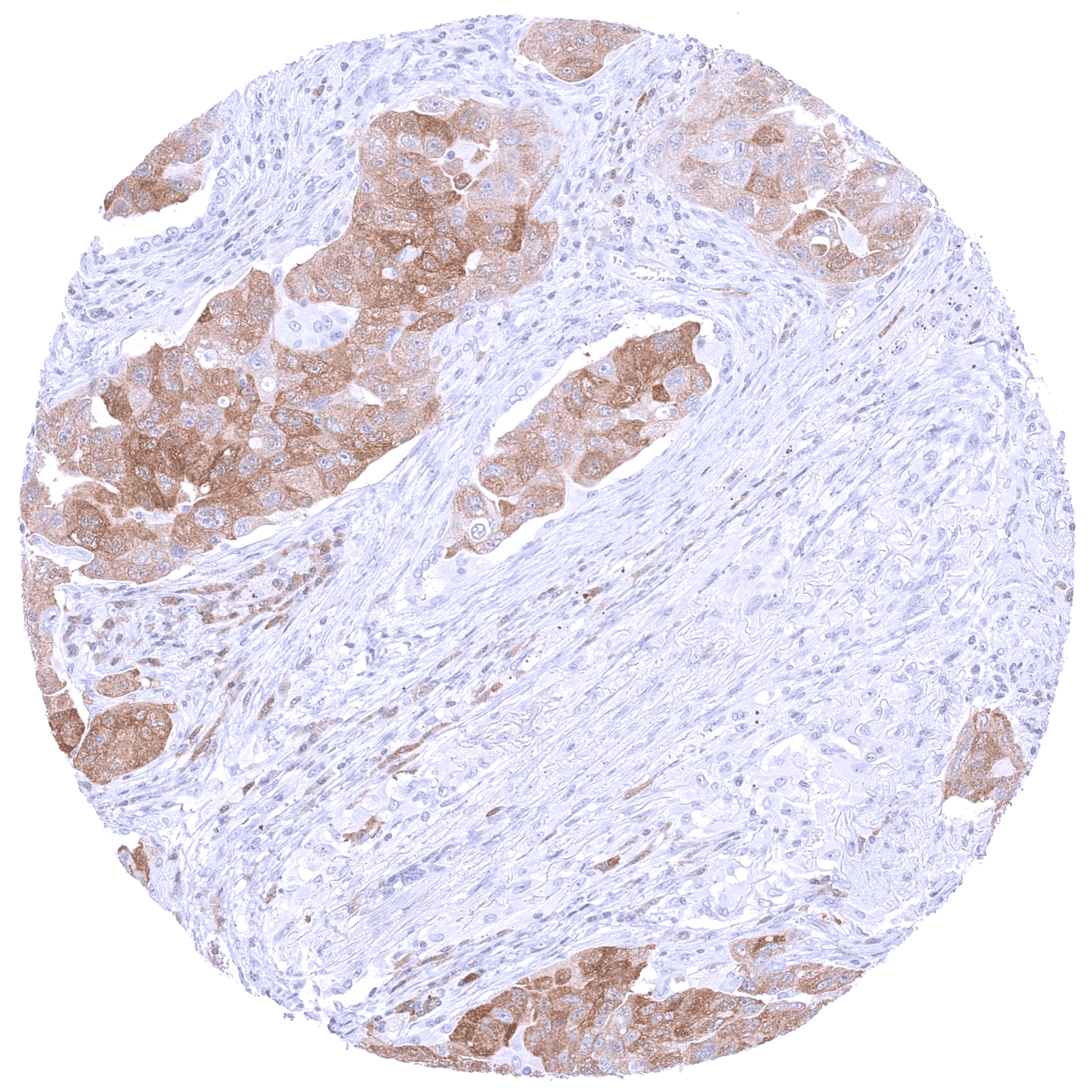
| Liver, Gallbladder, Pancreas | Liver | Weak, predominantly cytoplasmic PSAT1 staining of hepatocytes. | |
| Gallbladder | Negative | ||
| Pancreas | Cytoplasmic and nuclear PSAT1 staining of variable intensity of a subset of acinar cells. Islet cells are PSAT1 negative. | ||
| Kidney, urinary bladder | Kidney, cortex | Negative | |
| Kidney, medulla | Negative | ||
| Urinary bladder, urothelium | Negative | ||
| Kidney pelvis, mucosa | Negative | ||
| Male tissues | Prostate | Negative | |
| Seminal vesicle | Negative | ||
| Epididymis caput | Negative | ||
| Epididymis cauda | Negative | ||
| Testis | Weak to moderate cytoplasmic PSAT1 staining of Sertoli cells. | ||
| Female Tissues | Breast, glands | Negative | |
| Ectocervix | Negative | ||
| Endocervix | Negative | ||
| Endometrium, proliferation | Negative | ||
| Endometrium, secretion | Negative | ||
| Uterus, myometrium | Negative | ||
| Fallopian tube | Weak cytoplasmic and nuclear PSAT1 staining of a subset of cells. | ||
| Ovary, stroma | Negative | ||
| Ovary, follicular cyst | Negative | ||
| Ovary, corpus luteum | Negative | ||
| Amnion | Negative | ||
| Chorion | Negative | ||
| Amnion/Chorion | Negative | ||
| Placenta, early, decidua | Negative | ||
| Placenta, first trimenon | Negative | ||
| Placenta, mature | Negative | ||
| Muscle, connective & soft tissue | Aorta, intima | Negative | |
| Skeletal muscle | Negative | ||
| Aorta, media | Negative | ||
| Skeletal muscle, tongue | Negative | ||
| Heart, left ventricle | Negative | ||
| Kidney pelvis, muscular wall | Negative | ||
| Urinary bladder, muscular wall | Negative | ||
| Esophagus, muscular wall | Negative | ||
| Stomach, muscular wall | Negative | ||
| Ileum, muscular wall | Negative | ||
| Appendix, muscular wall | Negative | ||
| Colon descendens, muscular wall | Negative | ||
| Penis, glans, corpus spongiosum | Negative | ||
| Fat, white | Negative | ||
| Skin | Skin, surface | Negative | |
| Skin (hairs, sebaceous glands) | Negative | ||
| Anal canal, skin | Negative | ||
| Scrotum | Negative | ||
| Bone Marrow & lymphoid tissues | Bone marrow | Negative | |
| Thymus | Distinct nuclear and cytoplasmic PSAT1 staining of thymic epithelial cells. | ||
| Spleen | Negative | ||
| Lymph node | Negative | ||
| Tonsil, deep | Negative |
Details
More product details
More product details
Biology behind
Phosphoserine aminotransferase 1 (PSAT1) is coded by the PSAT1 gene at chromosome 9q21.2. It is a pivotal enzyme to produce serine and α-ketoglutarate, two critical metabolites for both carbon metabolism and the tricarboxylic acid cycle. PSAT1 is a rate limiting enzyme in the serine-glycine synthesis pathway which produces glycine, an essential nutrient for proliferating normal and neoplastic cells. Multiple studies have suggested that PSAT1 overexpression causes increased tumor cell proliferation, tumor progression, and poor patient prognosis in several different cancer types. Germ line PSAT1 mutations are among the causes for serine deficiency disorders which constitute inherited metabolic diseases with a broad phenotypic spectrum including the Neu–Laxova syndrome. In normal tissues, PSAT1 is mainly expressed in the brain, the kidney, and the pancreas but it also occurs in other cell types. Among tumors, PSAT1 expression is most often seen in brain, ovarian and endometrial carcinomas, although PSAT1 expression can also occur in tumors of various other organs.
Protocol Recommendations
Protocol Recommendations
Potential Research Applications
Potential Research Applications
Evidence For Antibody Specificity In I H C
Evidence For Antibody Specificity In I H C


