
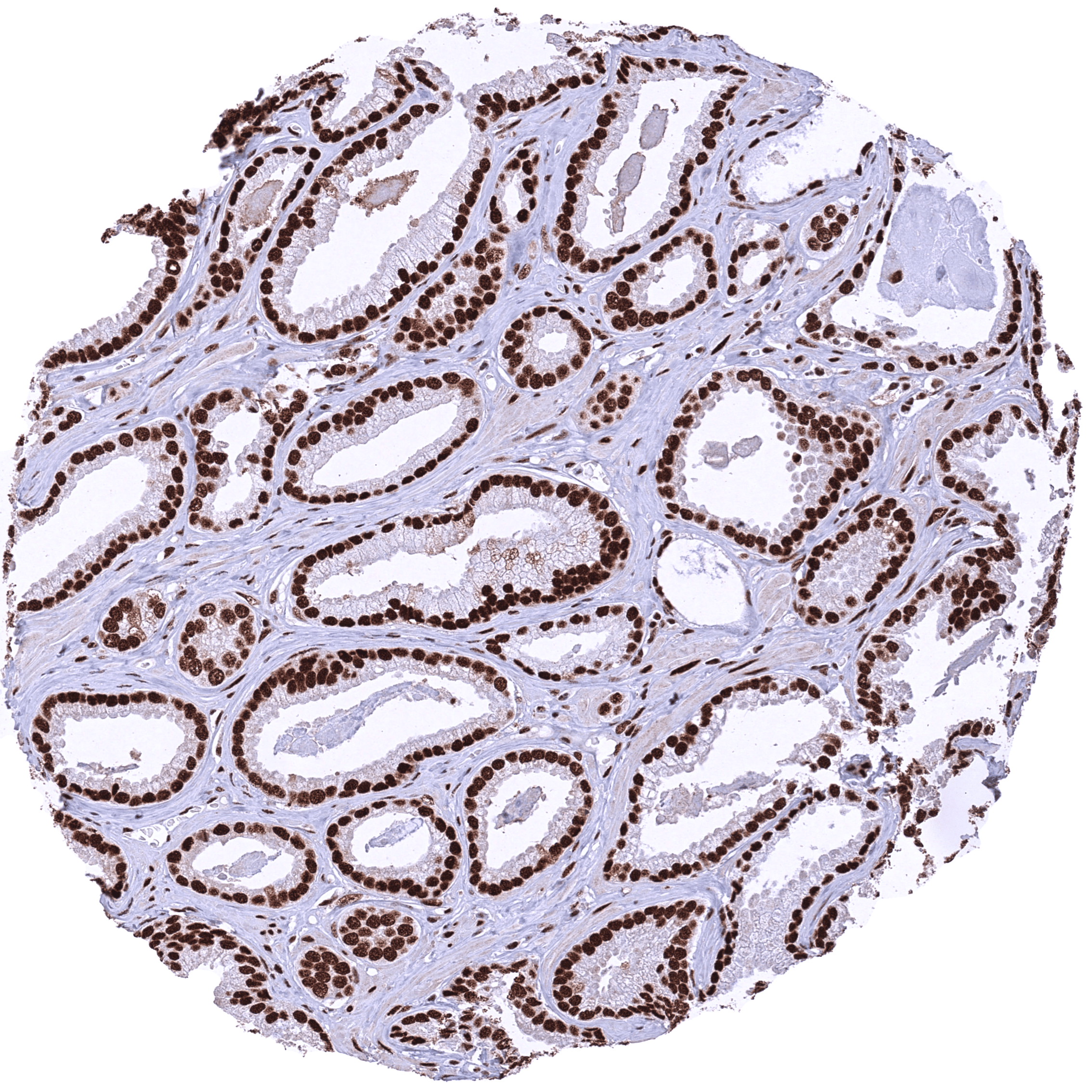
Breast gland with strong nuclear MRE11 staining of all cells. (Startbild)
Staining Pattern in Normal Tissues
Manual protocol
Freshly cut sections should be used (less than 10 days between cutting and staining). Heat-induced antigen retrieval for 5 minutes in an autoclave at 121°C in pH 7,8 Target Retrieval Solution buffer. Apply HMV328 at a dilution of 1:200 at 37°C for 60 minutes. Visualization of bound antibody by the EnVision Kit (Dako, Agilent) according to the manufacturer’s directions.
| Brain | Cerebrum, grey | Negative | |
| Cerebrum, white | Negative | ||
| Cerebellum, cortex | Negative | ||
| Cerebellum, white | Negative | ||
| Ganglion | Negative | ||
| Ependyma | Negative | ||
| Eye, retina | Negative | ||
| Endocrine Tissues | Thyroid | Strong nuclear MRE11 staining of all cells. | |
| Parathyroid gland | Strong nuclear MRE11 staining of all cells. | ||
| Adrenal gland | Distinct nuclear MRE11 staining of all cells. | ||
| Pituitary gland, anterior lobe | Negative | ||
| Pituitary gland, posterior lobe | Negative | ||
| Respiratory system | Lung bronchi | Negative | |
| Lung, bronchial glands | Negative | ||
| Nose, paranasal sinus | Negative | ||
| Lung, parenchyma | Negative | ||
| Proximal digestive tract | Lip | Negative | |
| Oral cavity | Negative | ||
| Tonsil, surface | Negative | ||
| Esophagus, mucosa | Negative | ||
| Lip, small salivary gland | Negative | ||
| Sublingual gland | Negative | ||
| Parotid gland | Negative | ||
| Submandibullary gland | Negative | ||
| Gastronintestinal tract | Stomach, antrum | Negative | |
| Stomach, fundus and corpus | Negative | ||
| Small intestine, duodenum | Negative | ||
| Duodenum, Brunner gland | Negative | ||
| Small intestine, ileum | Negative | ||
| Appendix | Strong nuclear MRE11 staining of all cells. | ||
| Colon descendens | Negative | ||
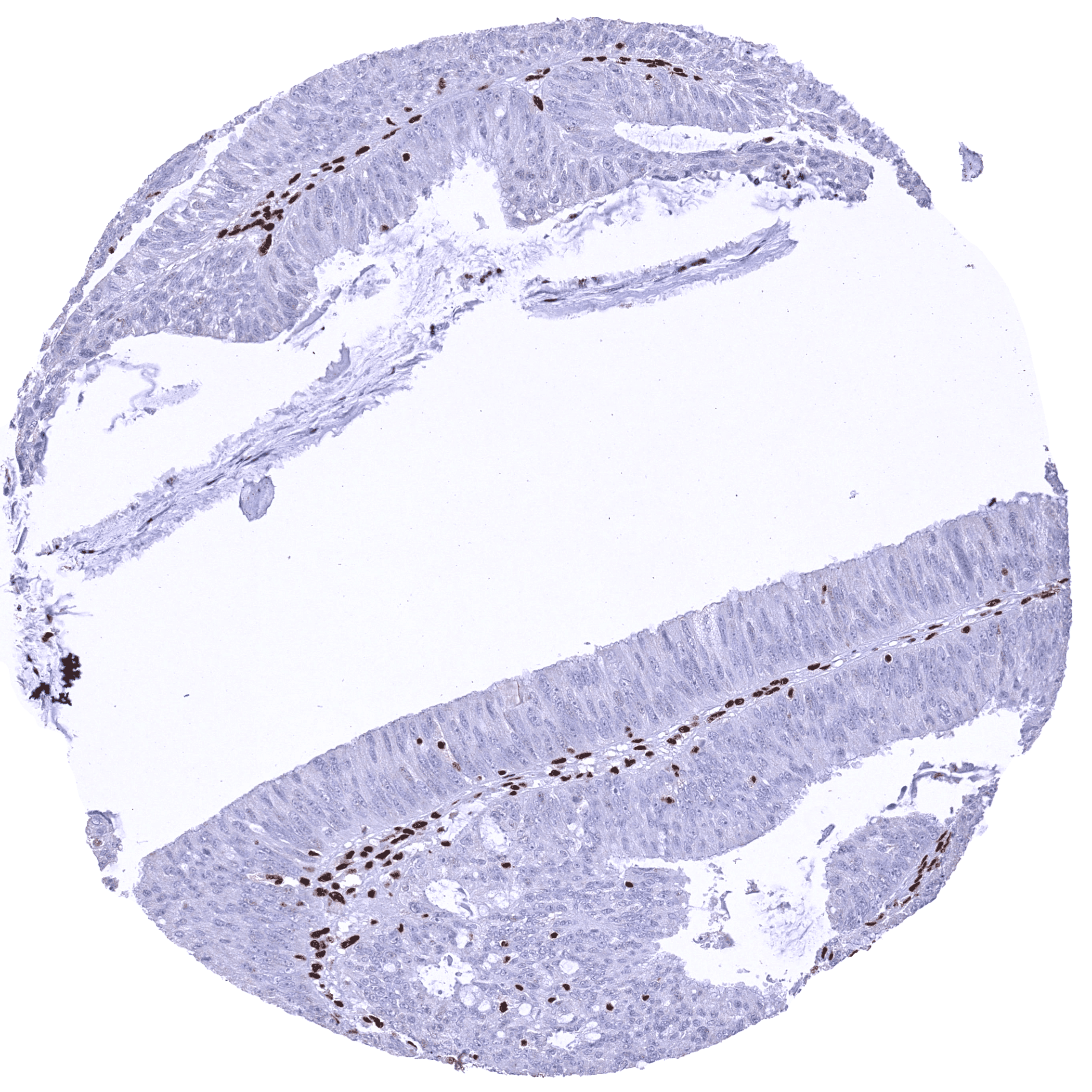
| Rectum | Distinct nuclear MRE11 staining of all cells. Slight but significant decrease of MRE11 staining from the crypt base to the superficial epithelial cell layers. | ||
| Anal canal, transition epithelium | Negative | ||
| Liver, Gallbladder, Pancreas | Liver | Nuclear MRE11 staining of most cells. Staining is clearly weakest in hepatocytes where it varies from negative to moderate. | |
| Gallbladder | Strong nuclear MRE11 staining of all cells. | ||
| Pancreas | Strong nuclear MRE11 staining of all cells. | ||
| Kidney, urinary bladder | Kidney, cortex | Negative | |
| Kidney, medulla | Negative | ||
| Urinary bladder, urothelium | Negative | ||
| Kidney pelvis, mucosa | Negative | ||
| Male tissues | Prostate | Strong nuclear MRE11 staining of all cells. | |
| Seminal vesicle | Negative | ||
| Epididymis caput | Negative | ||
| Epididymis cauda | Negative | ||
| Testis | Strong nuclear MRE11 staining of all cells. Additional cytoplasmic staining of one tubular cell type (Sertoli cells?). The staining intensity of germ cells decreases from spermatogonia to spermatids. | ||
| Female Tissues | Breast, glands | Negative | |
| Ectocervix | Negative | ||
| Endocervix | Negative | ||
| Endometrium, proliferation | Negative | ||
| Endometrium, secretion | Negative | ||
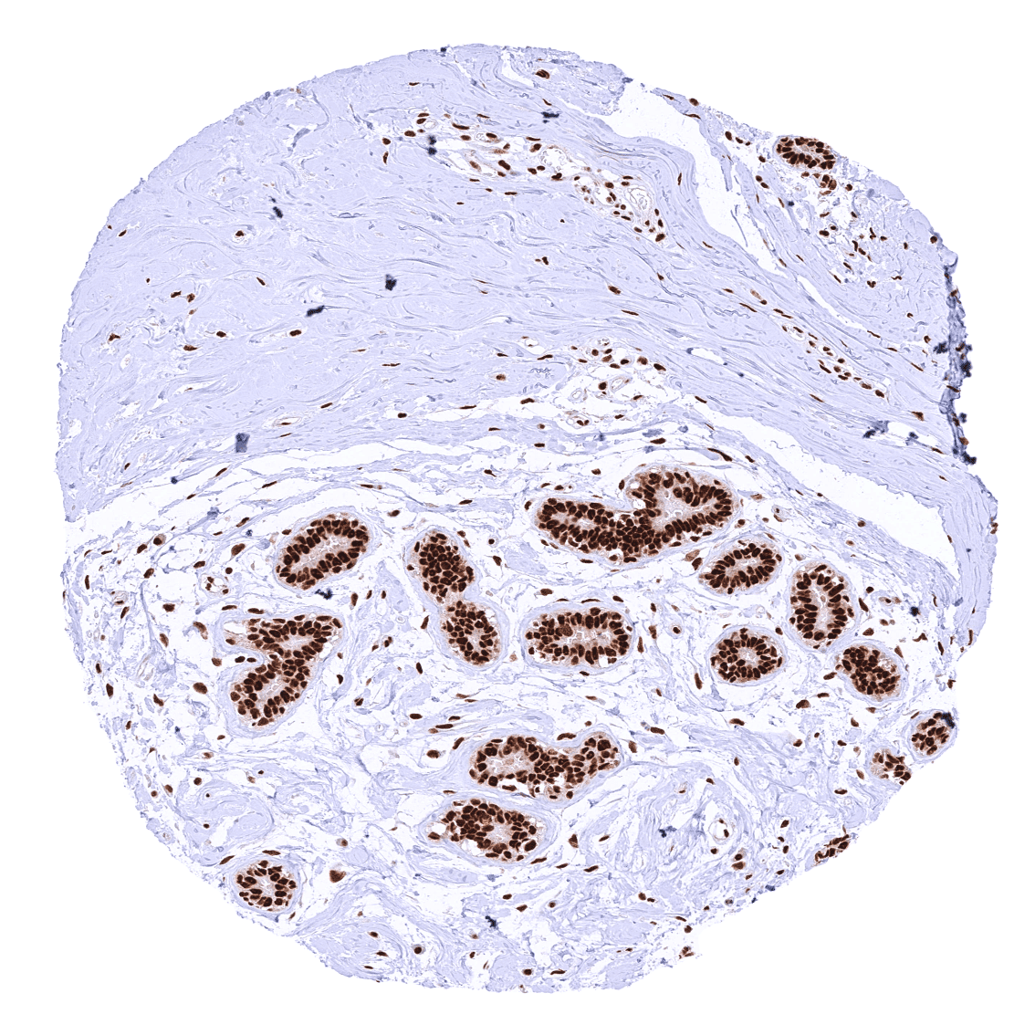
| Uterus, myometrium | Strong nuclear MRE11 staining of all cells. | ||
| Fallopian tube | Strong nuclear MRE11 staining of all cells. | ||
| Ovary, stroma | Negative | ||
| Ovary, follicular cyst | Negative | ||
| Ovary, corpus luteum | Negative | ||
| Amnion | Strong nuclear MRE11 staining of all cells. | ||
| Chorion | Strong nuclear MRE11 staining of all cells. | ||
| Amnion/Chorion | Negative | ||
| Placenta, early, decidua | Negative | ||
| Placenta, first trimenon | Negative | ||
| Placenta, mature | Nuclear MRE11 staining of all cells. However, the nuclear staining is somewhat weaker in syncytiotrophoblast cells than in all other cell types. | ||
| Muscle, connective & soft tissue | Aorta, intima | Negative | |
| Skeletal muscle | Strong nuclear MRE11 staining of all cells. | ||
| Aorta, media | Negative | ||
| Skeletal muscle, tongue | Negative | ||
| Heart, left ventricle | Negative | ||
| Kidney pelvis, muscular wall | Negative | ||
| Urinary bladder, muscular wall | Negative | ||
| Esophagus, muscular wall | Negative | ||
| Stomach, muscular wall | Negative | ||
| Ileum, muscular wall | Negative | ||
| Appendix, muscular wall | Negative | ||
| Colon descendens, muscular wall | Negative | ||
| Penis, glans, corpus spongiosum | Negative | ||
| Fat, white | Negative | ||
| Skin | Skin, surface | Negative | |
| Skin (hairs, sebaceous glands) | Negative | ||
| Anal canal, skin | Negative | ||
| Scrotum | Negative | ||
| Bone Marrow & lymphoid tissues | Bone marrow | Strong nuclear MRE11 staining of all cells. | |
| Thymus | Strong nuclear MRE11 staining of all cells. A few dispersed cells stand out because of a particularly strong nuclear MRE11 staining. | ||
| Spleen | Strong nuclear MRE11 staining of all cells. | ||
| Lymph node | Strong nuclear MRE11 staining of all cells. | ||
| Tonsil, deep | Negative |
Details
More product details
More product details
Biology behind
Double-strand break repair protein MRE11 (also named MRE11A) is an 80,6 kDa (predominant isoform) protein coded by the MRE11 gene on chromosome 11q21. Together with RAD50 and NBS1, MRE11 forms the MRN complex which is a key element in DNA damage response (DDR) and possesses both single-stranded DNA endonuclease and 3′ to 5′ exonuclease activities. The MRN complex is an early sensor for locating double strand DNA breaks (DSBs) and plays a direct role in both DSB repair and the recruiting of DDR proteins and activation of their downstream signaling. It regulates repair of DNA double-strand breaks in several contexts, including replication, telomere homeostasis, meiosis, apoptosis and immune system development. The MRN complex is involved in multiple different pathways of DSBs repair, including homologous recombination (HR), non-homologous end joining (NHEJ) and the (most error prone) pathway of microhomology-mediated end joining (MMEJ) repair. MRE11 is one of 6 enzymes required for MMEJ. MRE11 and the MRN complex are thought to play a key role in cancer radiosensitivity. MRE11 overexpressing cancers are generally considered to be more radioresistant and low-expressors are regarded as chemo-sensitive although this rule may not apply to all cancer types. Several MRE11 inhibitors are currently investigated in clinical trials as radiosensitizers.
Protocol Recommendations
Protocol Recommendations
Potential Research Applications
Potential Research Applications
Evidence For Antibody Specificity In I H C
Evidence For Antibody Specificity In I H C


