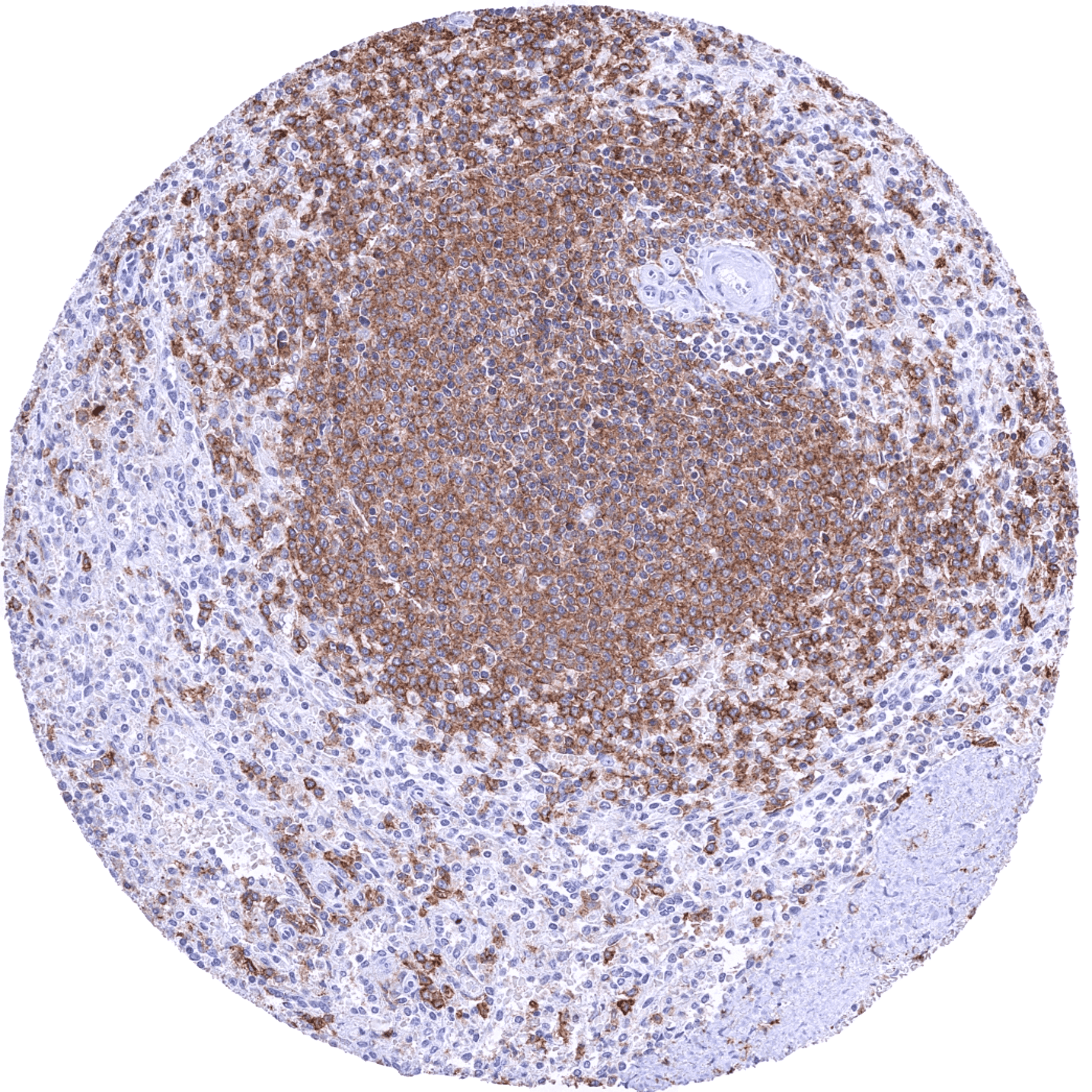
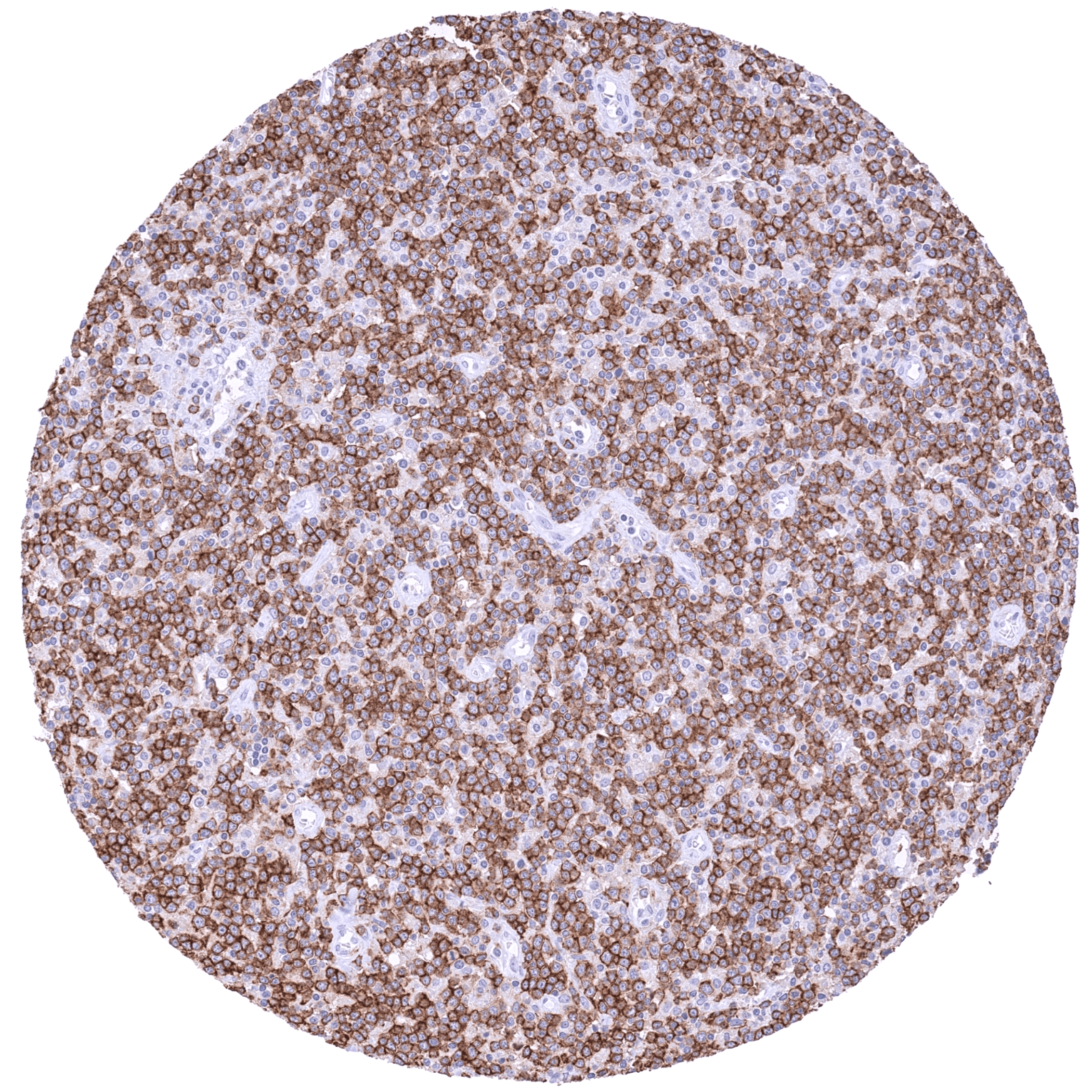
Staining Pattern in Normal Tissues
Manual protocol
Freshly cut sections should be used (less than 10 days between cutting and staining). Heat-induced antigen retrieval for 5 minutes in an autoclave at 121°C in pH 7,8 Target Retrieval Solution buffer. Apply HMV315 at a dilution of 1:200 at 37°C for 60 minutes. Visualization of bound antibody by the EnVision Kit (Dako, Agilent) according to the manufacturer’s directions.
Brain | Cerebrum | Negative | |
Cerebellum | Negative | ||
Endocrine tissues | Thyroid | Negative | |
Parathyroid | Negative | ||
Adrenal gland | Negative | ||
Pituitary gland | Distinct CD38B staining of some (monocytic and/or endothelial) cells, especially in the posterior lobe. | ||
Respiratory system | Respiratory epithelium | Negative | |
Lung | Negative | ||
Gastrointestinal tract | Salivary glands | Negative | |
Esophagus | Negative | ||
Stomach | Negative | ||
Duodenum | Negative | ||
Small intestine | Negative | ||
Appendix | Negative | ||
Colon | Negative | ||
Rectum | Negative | ||
Liver, Gallbladder, Pancreas | Liver | Moderate to strong CD38 staining of sinusoidal endothelium. | |
Gallbladder | Negative | ||
Pancreas | Negative | ||
Genitourinary | Kidney | Negative | |
Urothelium | Negative | ||
Male tissues | Prostate | Negative | |
Seminal Vesicles | Negative | ||
Testis | Negative | ||
Epididymis | Negative | ||
Female tissues | Breast | Negative | |
Uterus, myometrium | Negative | ||
Uterus, ectocervix | Negative | ||
Uterus, endocervix | Negative | ||
Uterus, endometrium | Negative | ||
Fallopian tube | Negative | ||
Ovary | Negative | ||
Placenta early | Strong CD32B positivity of all endothelial cells. | ||
Placenta mature | Strong CD32B positivity of all endothelial cells. | ||
Amnion | Negative | ||
Chorion | Negative | ||
Skin | Epidermis | Negative | |
Sebaceous glands | Negative | ||
Muscle, connective & soft tissues | Heart muscle | Negative | |
Skeletal muscle | Negative | ||
Smooth muscle | Negative | ||
Vessel walls | Endothelial cells of microvessels can be positive (only in selected tissue types). | ||
Fat | Negative | ||
Stroma | CD38B positivity occurs mainly in monocytic cells, B-lymphocytes, and may also be seen in the endothelium of microvessels (only in selected tissue types). Endothelial positivity is often difficult to discern from CD32B positive monocytic cell types. | ||
Endothelium | Endothelial cells of microvessels can stain CD32B positive in selected tissue types. | ||
Bone marrow & lymphoid tissues | Bone marrow | CD38B staining of a significant fraction of cells. | |
Lymph node | CD38B staining of variable intensity in a large fraction of B-lymphocytes and perhaps also other cells. CD32B staining is least intense in most germinal centre cells. | ||
Spleen | CD38B staining of variable intensity in most B-lymphocytes of the white pulpa. | ||
Thymus | CD38B staining of only few cells. | ||
Tonsil | CD38B staining of variable intensity in a large fraction of B-lymphocytes and perhaps also other cells. CD32B staining is least intense in most germinal centre cells. | ||
Remarks | *CD38B positivity of a fraction of inflammatory cells occurs in all tissues. In some tissues, CD32B positivity can also be seen in endothelia of microvessels. Endothelial positivity is often difficult to discern from CD32B positive monocytic cell types. |
Details
More product details
Biology behind
CD32B also termed FcγRIIB is an 40 kDa glycoprotein coded by the FCGR2B gene on chromosome 1q23.3. CD32B is a type I transmembrane receptor with low affinity for monomeric IgG. CD32B is the only known inhibitory Fc gamma receptor. It is expressed on subsets of lymphocytes, dendritic cells, and endothelial cells. CD32B reduces downstream signaling of co-localized activating receptors following ligand-induced crosslinking. CD32B is known as a critical regulatory element in B-cell homeostasis as it controls cell activation by counterbalancing the stimulatory activity of multiple receptors, including the B-cell antigen receptor (BCR). It down-regulates B cell activation by increasing the threshold for BCR activation and suppresses B cell-mediated Ag presentation to T cells. CD32B is of considerable therapeutic interest. On normal and malignant B cells, CD32B can play a role in internalizing the anti-CD20 antibody drug rituximab from the B cell surface which results in an abrogation of its cell-mediated anticancer mechanisms. Moreover, CD32B is involved in adaptation to all kinds of intravenous Ig treatments, and it can potentiate the immunostimulatory activities of certain therapeutic mAbs targeting TNFR family members. Therapeutic antibodies directly targeting CD32B are also under development. Suggested reading: https://jlb.onlinelibrary.wiley.com/doi/full/10.1002/jlb.2mir0917-354r.
Protocol Recommendations
Potential Research Applications
Evidence For Antibody Specificity In I H C