
Invasive urothelial carcinoma of the urinary bladder with complete absence of KDM6 staining in tumor cells.
Staining Pattern in Normal Tissues
Manual protocol
Freshly cut sections should be used (less than 10 days between cutting and staining). Heat-induced antigen retrieval for 5 minutes in an autoclave at 121°C in pH 7,8 Target Retrieval Solution buffer. Apply HMV311 at a dilution of 1:150 at 37°C for 60 minutes. Visualization of bound antibody by the EnVision Kit (Dako, Agilent) according to the manufacturer’s directions.
| Brain | Cerebrum, grey | Negative | |
| Cerebrum, white | Negative | ||
| Cerebellum, cortex | Negative | ||
| Cerebellum, white | Negative | ||
| Ganglion | Negative | ||
| Ependyma | Negative | ||
| Eye, retina | Negative | ||
| Endocrine Tissues | Thyroid | Moderate to strong nuclear KDM6A staining of follicular epithelial cells. | |
| Parathyroid gland | Moderate to strong nuclear KDM6A staining of epithelial cells. | ||
| Adrenal gland | Moderate to strong nuclear KDM6A staining of adrenocortical cells. | ||
| Pituitary gland, anterior lobe | Moderate to strong nuclear KDM6A staining of epithelial cells | ||
| Pituitary gland, posterior lobe | Faint nuclear KDM6A staining of pituicytes | ||
| Respiratory system | Lung bronchi | Negative | |
| Lung, bronchial glands | Moderate to strong nuclear KDM6A staining of respiratory epithelial cells. | ||
| Nose, paranasal sinus | Negative | ||
| Lung, parenchyma | Negative | ||
| Proximal digestive tract | Lip | Negative | |
| Oral cavity | Negative | ||
| Tonsil, surface | Negative | ||
| Esophagus, mucosa | Negative | ||
| Lip, small salivary gland | Negative | ||
| Sublingual gland | Negative | ||
| Parotid gland | Moderate to strong nuclear KDM6A staining of all epithelial cells | ||
| Submandibullary gland | Negative | ||
| Gastronintestinal tract | Stomach, antrum | Negative | |
| Stomach, fundus and corpus | Negative | ||
| Small intestine, duodenum | Negative | ||
| Duodenum, Brunner gland | Moderate to strong nuclear KDM6A staining of Brunner gland cells. | ||
| Small intestine, ileum | Negative | ||
| Appendix | Strong nuclear KDM6A staining of epithelial and inflammatory cells. | ||
| Colon descendens | Negative | ||
| Rectum | Negative | ||
| Anal canal, transition epithelium | Negative | ||
| Liver, Gallbladder, Pancreas | Liver | Moderate nuclear KDM6A staining of hepatocytes. Stronger staining of other cell types. | |
| Gallbladder | Strong nuclear KDM6A staining of epithelial cells. | ||
| Pancreas | Moderate to strong nuclear KDM6A staining of acinar cells. Stronger staining of ductal and islet cells. | ||
| Kidney, urinary bladder | Kidney, cortex | Moderate nuclear KDM6A staining of epithelial cells of all types. | |
| Kidney, medulla | Moderate nuclear KDM6A staining of collecting ducts cells. | ||
| Urinary bladder, urothelium | Strong nuclear KDM6A staining of urothelial cells. | ||
| Kidney pelvis, mucosa | Negative | ||
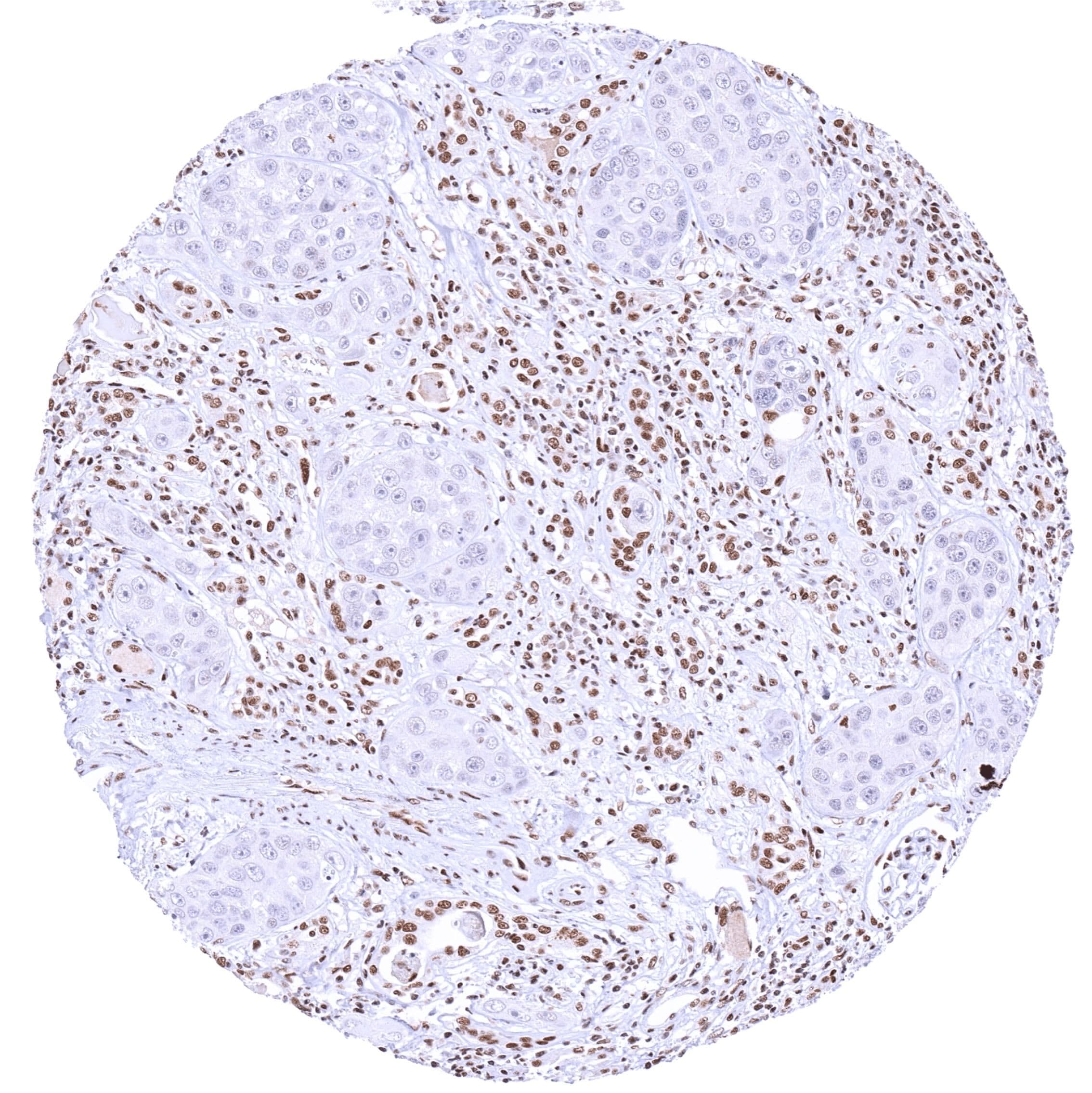
| Male tissues | Prostate | Moderate nuclear KDM6A staining of all epithelial cells. | |
| Seminal vesicle | Strong nuclear KDM6A staining of epithelial cells. | ||
| Epididymis caput | Moderate to strong nuclear KDM6A staining of epithelial cells of the caput. Staining is less intense in basal cells. | ||
| Epididymis cauda | Moderate to strong nuclear KDM6A staining of epithelial cells of the cauda. Staining is less intense in basal cells. | ||
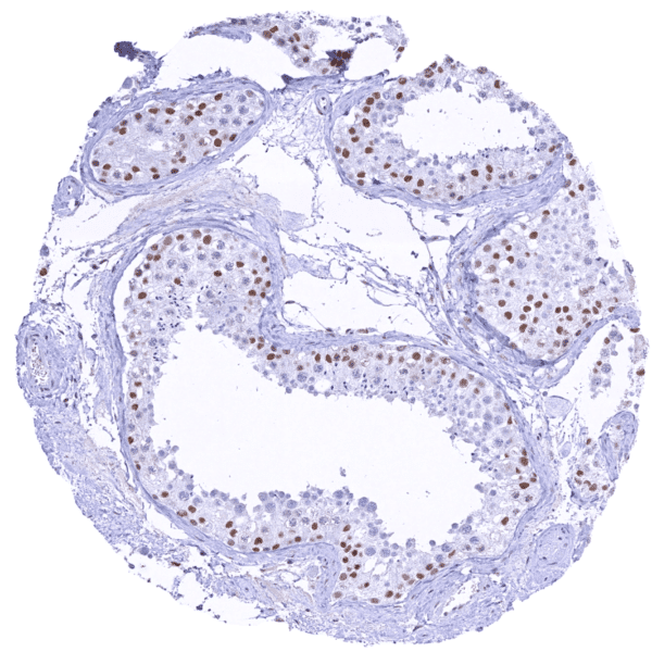
| Testis | Moderate to strong nuclear KDM6A staining of Sertoli cells. KDM6A staining is weak in spermatogonia and virtually absent in more mature cells of spermatogenesis. | ||
| Female Tissues | Breast, glands | Strong nuclear KDM6A staining of basal and luminal cells of breast glands. | |
| Ectocervix | Moderate to strong nuclear KDM6A staining of squamous epithelial cells. Staining is stronger in the lower than in the upper cell layers. | ||
| Endocervix | Strong nuclear KDM6A staining of epithelial cells. | ||
| Endometrium, proliferation | Negative | ||
| Endometrium, secretion | Negative | ||
| Uterus, myometrium | Moderate to strong nuclear KDM6A staining of muscular cells. | ||
| Fallopian tube | Strong nuclear KDM6A staining of all epithelial cells. | ||
| Ovary, stroma | Moderate nuclear KDM6A staining of stroma cells. | ||
| Ovary, follicular cyst | Negative | ||
| Ovary, corpus luteum | Strong nuclear KDM6A staining of corpus luteum cells. | ||
| Amnion | Weak nuclear KDM6A staining of amnion cells. | ||
| Chorion | Moderate to strong staining of chorion cells. | ||
| Amnion/Chorion | Negative | ||
| Placenta, early, decidua | Strong nuclear KDM6A staining of trophoblastic cells, stroma cells and endothelial cells. | ||
| Placenta, first trimenon | Negative | ||
| Placenta, mature | Strong nuclear KDM6A staining of trophoblastic cells, stroma cells and endothelial cells. | ||
| Muscle, connective & soft tissue | Aorta, intima | Negative | |
| Skeletal muscle | Moderate nuclear KDM6A staining of myocytes. | ||
| Aorta, media | Weak nuclear KDM6A staining of cells. | ||
| Skeletal muscle, tongue | Negative | ||
| Heart, left ventricle | Negative | ||
| Kidney pelvis, muscular wall | Negative | ||
| Urinary bladder, muscular wall | Weak to moderate nuclear KDM6A staining of muscular cells. | ||
| Esophagus, muscular wall | Negative | ||
| Stomach, muscular wall | Negative | ||
| Ileum, muscular wall | Negative | ||
| Appendix, muscular wall | Weak to moderate nuclear KDM6A staining of muscular and ganglion cells. | ||
| Colon descendens, muscular wall | Weak to moderate nuclear KDM6A staining of muscular cells. | ||
| Penis, glans, corpus spongiosum | Negative | ||
| Fat, white | Faint nuclear KDM6A staining of some fat cells. | ||
| Skin | Skin, surface | Negative | |
| Skin (hairs, sebaceous glands) | Negative | ||
| Anal canal, skin | Negative | ||
| Scrotum | Negative | ||
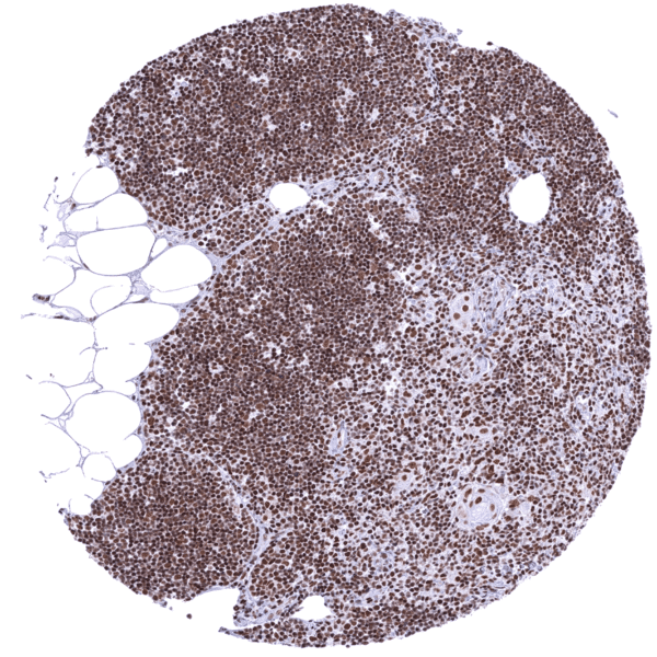
| Bone Marrow & lymphoid tissues | Bone marrow | Strong nuclear KDM6A staining of virtually all cells. | |
| Thymus | Intense nuclear KDM6A staining of all cell types. | ||
| Spleen | Moderate to strong staining of all cell types. | ||
| Lymph node | Moderate to strong staining of lymphocytes and other cell types. | ||
| Tonsil, deep | Negative |
KDM6A
(HMV311)
KDM6A is an Important enzyme for differentiation of embryonic stem cells and development of various tissues.
Details
More product details
More product details
Biology behind
The lysine-specific demethylase 6A (KDM6A), also termed “Ubiquitously transcribed tetratricopeptide repeat on chromosome X” (UTX) is a 154 kDa protein coded by the KDM6A gene on chromosome Xp11.3. The gene is 5,438 bp long and contains 29 exons. It belongs to a family of Jumonji (JmjC) domain-containing enzymes which impact the demethylation of H3K27me2/3. H3K27me2/3 indicates the 27th amino acid on histone H3 which is known to represent a repressive histone modification. KDM6A regulates the transcription and expression of downstream genes, thereby regulating cell fate and functional cell characteristics. Depending on the molecular context, KDM6A can either promote or suppress cell growth. In evolving tissues, KDM6A is implicated in myogenesis, cardiac development, pancreas development, neural stem cell differentiation and immune cell functions. KDM6A is often mutated in various cancer types including bladder cancer, plasmacytoma, renal cell carcinoma, prostate cancer, and others. Mutations of KDM6A are a cause for Kabuki syndrome, a rare congenital anomaly syndrome characterized by intellectual disability, growth retardation, and multiple congenital abnormalities.
Protocol Recommendations
Protocol Recommendations
Potential Research Applications
Potential Research Applications
Evidence For Specificity In I H C
Evidence For Specificity In I H C


