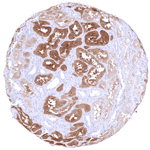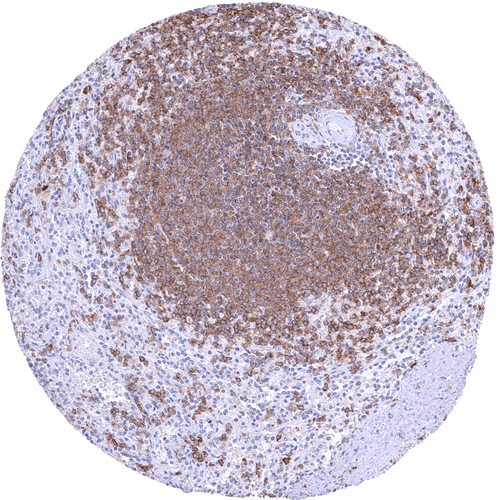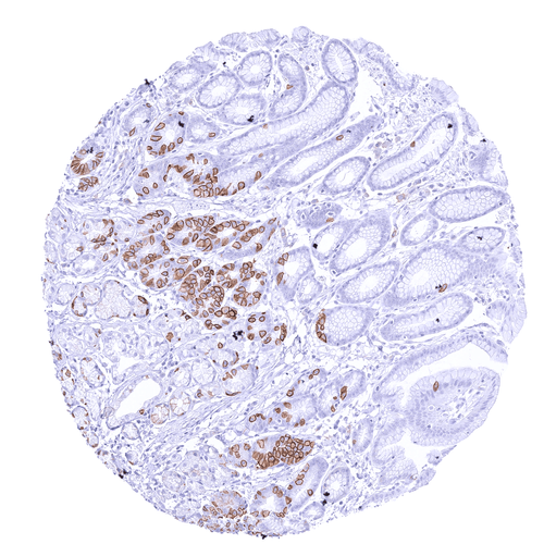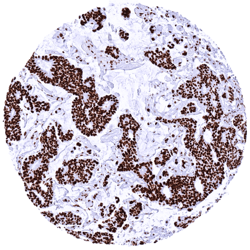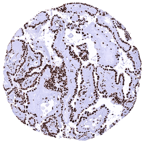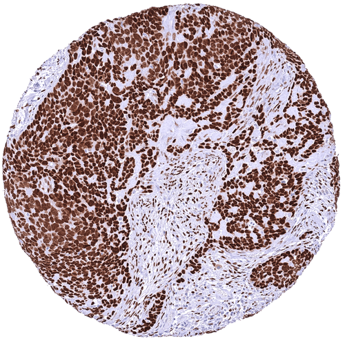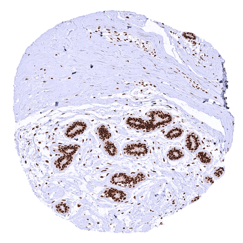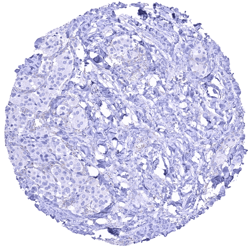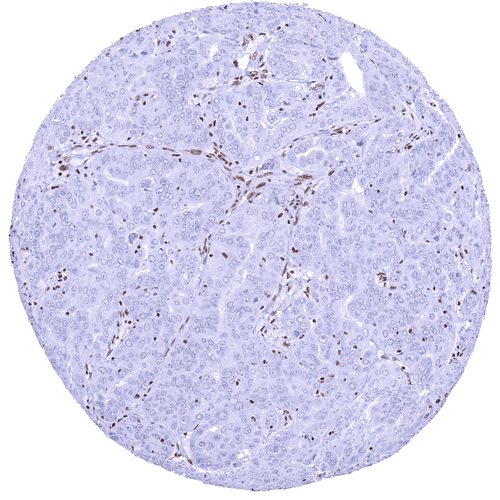

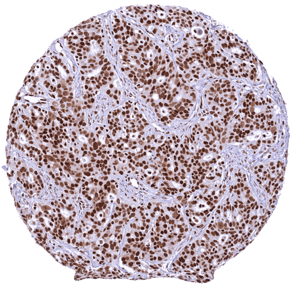
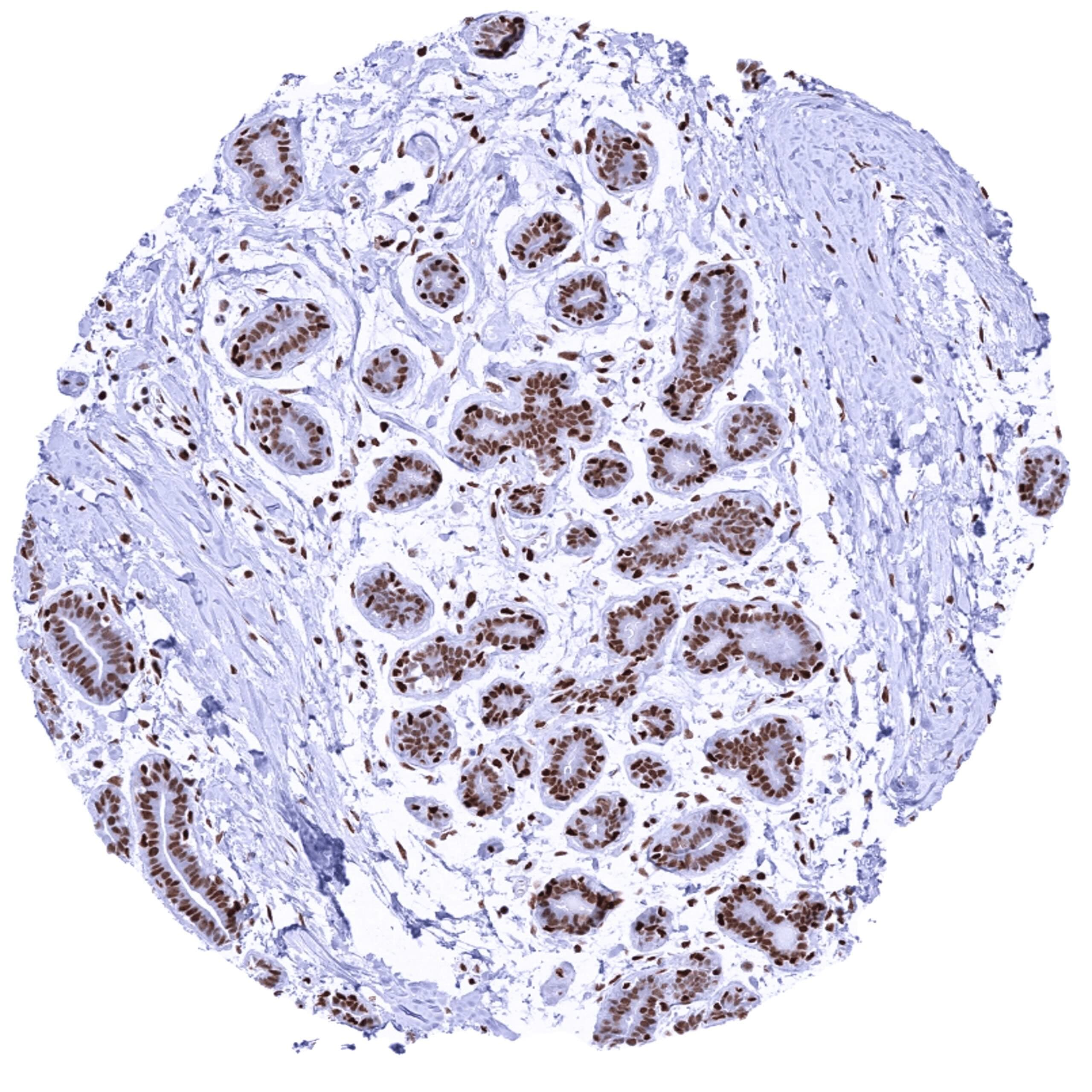
Breast glands showing a moderate to strong GR positivity of myoepithelial and luminal cells.
Staining Pattern in Normal Tissues
Manual protocol
Freshly cut sections should be used (less than 10 days between cutting and staining). Heat-induced antigen retrieval for 5 minutes in an autoclave at 121°C in pH 7,8 Target Retrieval Solution buffer. Apply HMV304 at a dilution of 1:150 at 37°C for 60 minutes. Visualization of bound antibody by the EnVision Kit (Dako, Agilent) according to the manufacturer’s directions.
| Brain | Cerebrum, grey | Moderate GR positivity of neuronal cells. | |
| Cerebrum, white | Negative | ||
| Cerebellum, cortex | Absence of unequivocal GR staining (perhaps due to overfixation of tissue) | ||
| Cerebellum, white | Negative | ||
| Ganglion | Negative | ||
| Ependyma | Negative | ||
| Eye, retina | Negative | ||
| Endocrine Tissues | Thyroid | Moderate to strong GR positivity of follicular epithelial cells. | |
| Parathyroid gland | Moderate to strong GR positivity of epithelial cells. | ||
| Adrenal gland | Moderate to strong GR positivity of medullary cells while adrenocortical cells are largely GR negative. | ||
| Pituitary gland, anterior lobe | Moderate to strong GR positivity of epithelial cells. Weak GR positivity of pituicytes. | ||
| Pituitary gland, posterior lobe | Weak GR positivity of pituicytes | ||
| Respiratory system | Lung bronchi | Moderate to strong GR positivity of respiratory epithelial cells. | |
| Lung, bronchial glands | Strong GR positivity of pneumocytes. | ||
| Nose, paranasal sinus | Negative | ||
| Lung, parenchyma | Negative | ||
| Proximal digestive tract | Lip | Negative | |
| Oral cavity | Negative | ||
| Tonsil, surface | Negative | ||
| Esophagus, mucosa | Intense GR staining of squamous epithelial cells. Staining intensity decreases sligthly towards the surface. | ||
| Lip, small salivary gland | Negative | ||
| Sublingual gland | Negative | ||
| Parotid gland | Negative | ||
| Submandibullary gland | Moderate to strong GR positivity of all epithelial cells. | ||
| Gastronintestinal tract | Stomach, antrum | Moderate GR positivity of superficial epithelial cells. Staining is weakest in gastric glands. | |
| Stomach, fundus and corpus | Moderate GR positivity of superficial epithelial cells. Staining is weakest in gastric glands. | ||
| Small intestine, duodenum | Moderate GR positivity of epithelial cells. GR staining is stronger in lymphocytes. | ||
| Duodenum, Brunner gland | Strong GR positivity of superficial epithelial cells. Staining is somewhat weaker in the crypts.Moderate GR positivity of Brunner gland cells. | ||
| Small intestine, ileum | Negative | ||
| Appendix | Moderate GR positivity of superficial epithelial cells. Epithelial cell staining decreases markedly towards the base of crypts where it can be absent in a fraction of cells. GR staining is markedly stronger in lymphocytes. | ||
| Colon descendens | Moderate GR positivity of epithelial cells. | ||
| Rectum | Moderate GR positivity of superficial epithelial cells. Epithelial cell staining decreases markedly towards the base of crypts where it can be absent in a fraction of cells. GR staining is markedly stronger in lymphocytes. | ||
| Anal canal, transition epithelium | Negative | ||
| Liver, Gallbladder, Pancreas | Liver | Moderate GR positivity of hepatocytes, ductal cells and of lymphocytes. | |
| Gallbladder | Strong GR positivity of epithelial cells. | ||
| Pancreas | Moderate to strong GR positivity of acinar cells while islet cells show a strong staining. | ||
| Kidney, urinary bladder | Kidney, cortex | Moderate to strong GR positivity of all epithelial cells. | |
| Kidney, medulla | Moderate to strong GR positivity of all epithelial cells. | ||
| Urinary bladder, urothelium | Weak to moderate GR positivity of the basal and suprabasal cell layers of the urohelium. Superficial cell layers are largely GR negative. | ||
| Kidney pelvis, mucosa | Weak to moderate GR positivity of the basal and suprabasal cell layers of the urohelium. Superficial cell layers are largely GR negative. | ||
| Male tissues | Prostate | Moderate to strong GR positivity of epithelial cells. | |
| Seminal vesicle | Moderate to strong GR positivity of all epithelial cells. | ||
| Epididymis caput | Moderate to strong GR positivity of all epithelial cells of the caput epididymis. | ||
| Epididymis cauda | Negative | ||
| Testis | Intense GR positivity of Sertoli and Leydig cells. Cells of the spermatogenesis do not show significant GR staining. | ||
| Female Tissues | Breast, glands | Moderate to strong GR positivity of myoepithelial and luminal cells.s | |
| Ectocervix | Significant GR staining of squamous epithelial cells. Staining intensity decreases sligthly towards the surface. | ||
| Endocervix | Moderate GR positivity of epithelial cells. | ||
| Endometrium, proliferation | Negative | ||
| Endometrium, secretion | Moderate GR positivity of stroma cells while endometrium cells are largely GR negative. | ||
| Uterus, myometrium | Strong GR positivity of myometrial cells. | ||
| Fallopian tube | Strong GR positivity of all epithelial cells. | ||
| Ovary, stroma | Strong GR positivity of stromal cells. | ||
| Ovary, follicular cyst | Granulosa cells are largely GR negative. | ||
| Ovary, corpus luteum | Moderate to strong GR positivity of corpus luteum cells. | ||
| Amnion | Negative | ||
| Chorion | Negative | ||
| Amnion/Chorion | Strong GR positivity of amnion and chorion cells. | ||
| Placenta, early, decidua | Strong GR staining of trophoblast and stroma cells. | ||
| Placenta, first trimenon | Negative | ||
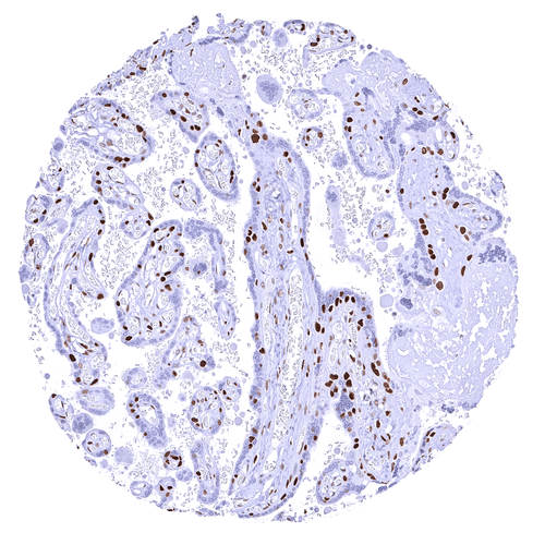
| Placenta, mature | Strong GR staining of stroma cells while trophoblast cells are negative. Moderate GR positivity of decidua cells. | ||
| Muscle, connective & soft tissue | Aorta, intima | Negative | |
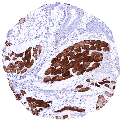
| Skeletal muscle | Strong GR positivity of skeletal muscle cells. | ||
| Aorta, media | Moderate to strong GR positivity of spindle shaped cells in the media. | ||
| Skeletal muscle, tongue | Negative | ||
| Heart, left ventricle | Negative | ||
| Kidney pelvis, muscular wall | Negative | ||
| Urinary bladder, muscular wall | Negative | ||
| Esophagus, muscular wall | Negative | ||
| Stomach, muscular wall | Negative | ||
| Ileum, muscular wall | Negative | ||
| Appendix, muscular wall | Negative | ||
| Colon descendens, muscular wall | Negative | ||
| Penis, glans, corpus spongiosum | Negative | ||
| Fat, white | Strong GR positivity of fat cells. | ||
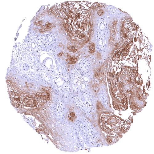
| Skin | Skin, surface | Intense GR positivity of squamous epithelial cells. Staining intensity decreases slightly towards the surface. | |
| Skin (hairs, sebaceous glands) | Negative | ||
| Anal canal, skin | Negative | ||
| Scrotum | Negative | ||
| Bone Marrow & lymphoid tissues | Bone marrow | Strong GR positivity of almost all hematopoetic cells. | |
| Thymus | Strong GR positivity of lymphocytic cells. GR staining is only weak in squamous epithelial cells of corpuscles of Hassall‘s. | ||
| Spleen | Negative | ||
| Lymph node | Negative | ||
| Tonsil, deep | Strong GR positivity of lymphocytic cells.Intense GR positivity of squamous epithelial cells with somewhat decreasing intensity towards the surface. |
Glucocorticoid Receptor
(HMV304)
Glucocorticoid Receptor is a pivotal multifunctional nuclear receptor protein.
Details
More product details
More product details
Biology behind
The glucocorticoid receptor (GR, or GCR) is a 777-amino acid multidomain nuclear protein which is coded by the NR3C1 (nuclear receptor subfamily 3, group C, member 1) gene at 5q31 which consists of ten exons (1 to 9β). Together with the other steroid receptors progesterone receptor (PR), androgen receptor (AR), the estrogen receptors (ERα and ERβ), and the mineralocorticoid receptor (MR), GR builds the nuclear receptor (NR) superfamily subgroup 3. GR is the receptor for cortisol and other glucocorticoids. It is expressed in most human cell types and regulates the transcription of thousands of genes involved in metabolism, development, stress and inflammatory responses. In the absence of its ligand, GR is bound to heat shock proteins in the cytoplasm. Ligand binding results in GR dislocation to the nucleus, binding to specific glucocorticoid response elements (GREs), recruitment of context specific transcriptional coregulators, and consequently, either activation or repression of the respective target genes. GR coregulators include a large and diverse group of proteins such as histone modifying enzymes, histone acetyltransferases or deacetylases, and many others. GR and glucocorticoids have a critical impact on a very broad spectrum of physiological processes. For example, GR appears to be an important adaptor for endocrine influence – specifically the stress response – on the brain and may play a critical role in psychological disorders such as depression and post-traumatic stress disorder. GR expression also plays a complex role in tumorigenesis which goes beyond its effects on the immune system. In several tumor types, both an oncogenic and a tumor suppressive function of GR has been found depending on specific tumor conditions.
Protocol Recommendations
Protocol Recommendations
Potential Research Applications
Potential Research Applications
Evidence For Specificity In I H C
Evidence For Specificity In I H C