

Strong TYMS staining of a large fraction of thymic lymphocytes.
Staining Pattern in Normal Tissues
Manual protocol
Freshly cut sections should be used (less than 10 days between cutting and staining). Heat-induced antigen retrieval for 5 minutes in an autoclave at 121°C in pH 7,8 Target Retrieval Solution buffer. Apply HMV305 at a dilution of 1:150 at 37°C for 60 minutes. Visualization of bound antibody by the EnVision Kit (Dako, Agilent) according to the manufacturer’s directions.
| Brain | Cerebrum, grey | Negative | |
| Cerebrum, white | Negative | ||
| Cerebellum, cortex | Negative | ||
| Cerebellum, white | Negative | ||
| Ganglion | Negative | ||
| Ependyma | Negative | ||
| Eye, retina | Negative | ||
| Endocrine Tissues | Thyroid | Negative | |
| Parathyroid gland | Negative | ||
| Adrenal gland | Negative | ||
| Pituitary gland, anterior lobe | Negative | ||
| Pituitary gland, posterior lobe | Negative | ||
| Respiratory system | Lung bronchi | Negative | |
| Lung, bronchial glands | Negative | ||
| Nose, paranasal sinus | Negative | ||
| Lung, parenchyma | Negative | ||
| Proximal digestive tract | Lip | Negative | |
| Oral cavity | Negative | ||
| Tonsil, surface | Negative | ||
| Esophagus, mucosa | Negative | ||
| Lip, small salivary gland | Negative | ||
| Sublingual gland | Negative | ||
| Parotid gland | Negative | ||
| Submandibullary gland | Negative | ||
| Gastronintestinal tract | Stomach, antrum | Weak to moderate, nuclear and cytoplasmic TYMS staining of a fraction of epithelial cells. | |
| Stomach, fundus and corpus | Weak to moderate, nuclear and cytoplasmic TYMS staining of a fraction of epithelial cells. | ||
| Small intestine, duodenum | Negative | ||
| Duodenum, Brunner gland | Negative | ||
| Small intestine, ileum | Negative | ||
| Appendix | Weak to moderate, nuclear and cytoplasmic TYMS staining of a fraction of crypt epithelial cells while staining is strong in many lymphocytic cells | ||
| Colon descendens | Faint, nuclear and cytoplasmic TYMS staining of a fraction of crypt epithelial cells | ||
| Rectum | Weak to moderate, nuclear and cytoplasmic TYMS staining of a large subset of crypt epithelial cells. | ||
| Anal canal, transition epithelium | Negative | ||
| Liver, Gallbladder, Pancreas | Liver | Negative | |
| Gallbladder | Weak to moderate, nuclear and cytoplasmic TYMS staining of a subset of epithelial cells. | ||
| Pancreas | Weak to moderate, nuclear and cytoplasmic TYMS staining of very few epithelial cells. | ||
| Kidney, urinary bladder | Kidney, cortex | Negative | |
| Kidney, medulla | Negative | ||
| Urinary bladder, urothelium | Negative | ||
| Kidney pelvis, mucosa | Negative | ||
| Male tissues | Prostate | Negative | |
| Seminal vesicle | Negative | ||
| Epididymis caput | Negative | ||
| Epididymis cauda | Negative | ||
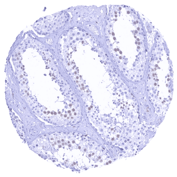
| Testis | Weak to moderate, nuclear and cytoplasmic TYMS staining of spermatocytes. | ||
| Female Tissues | Breast, glands | Strong, nuclear and cytoplasmic TYMS staining of a subset of luminal epithelial cells of breast glands. | |
| Ectocervix | Negative | ||
| Endocervix | Negative | ||
| Endometrium, proliferation | Negative | ||
| Endometrium, secretion | Negative | ||
| Uterus, myometrium | Negative | ||
| Fallopian tube | Negative | ||
| Ovary, stroma | Negative | ||
| Ovary, follicular cyst | Negative | ||
| Ovary, corpus luteum | Moderate to strong, nuclear and cytoplasmic TYMS staining of some endothelial cells. | ||
| Amnion | Negative | ||
| Chorion | Negative | ||
| Amnion/Chorion | Negative | ||
| Placenta, early, decidua | Variable, weak to strong, nuclear and cytoplasmic TYMS staining of few cells (cytotrophoblast, stromal) | ||
| Placenta, first trimenon | Negative | ||
| Placenta, mature | Variable, weak to strong, nuclear and cytoplasmic TYMS staining of few cells (trophoblast, endothelial) | ||
| Muscle, connective & soft tissue | Aorta, intima | Negative | |
| Skeletal muscle | Negative | ||
| Aorta, media | Negative | ||
| Skeletal muscle, tongue | Negative | ||
| Heart, left ventricle | Negative | ||
| Kidney pelvis, muscular wall | Negative | ||
| Urinary bladder, muscular wall | Negative | ||
| Esophagus, muscular wall | Negative | ||
| Stomach, muscular wall | Negative | ||
| Ileum, muscular wall | Negative | ||
| Appendix, muscular wall | Negative | ||
| Colon descendens, muscular wall | Negative | ||
| Penis, glans, corpus spongiosum | Negative | ||
| Fat, white | Negative | ||
| Skin | Skin, surface | Negative | |
| Skin (hairs, sebaceous glands) | Negative | ||
| Anal canal, skin | Negative | ||
| Scrotum | Negative | ||
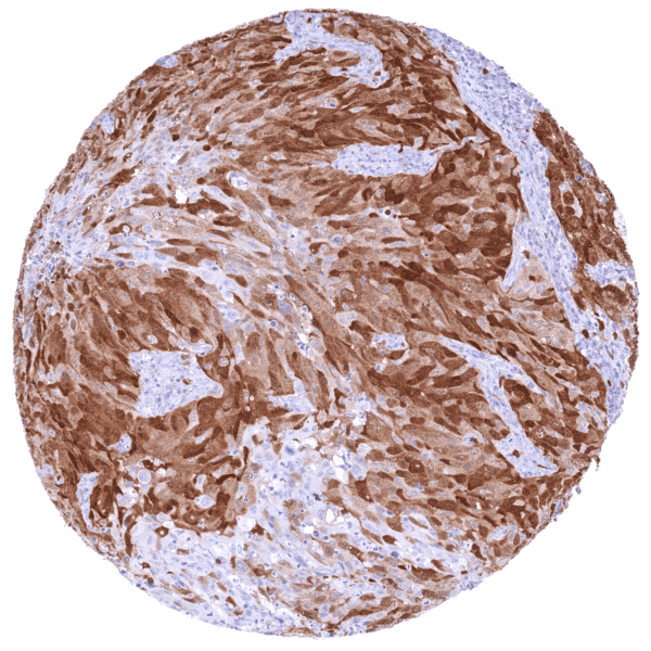
| Bone Marrow & lymphoid tissues | Bone marrow | Moderate to strong, nuclear and cytoplasmic TYMS staining of a large subset of hematopoetic cells. | |
| Thymus | Strong TYMS staining of a large fraction of lymphocytes. | ||
| Spleen | Moderate to strong TYMS staining of a fraction of lymphocytic cells. | ||
| Lymph node | Strong TYMS staining of a fraction of lymphocytic cells. | ||
| Tonsil, deep | Variable, weak to strong, nuclear and cytoplasmic TYMS staining of a fraction of lymphocytes, especially in the germinal centre. Weak to moderate, predominantly nuclear TYMS staining of a subset of suprabasal squamous epithelial cells. |
Details
More product details
More product details
Biology behind
Thymidylate synthase (TS, TYMS) is a 32-35kD enzyme which is coded by the TYMS gene at 18p11.32. TYMS catalyzes the conversion of deoxyuridine monophosphate (dUMP) to deoxythymidine monophosphate (dTMP) which is one of the nucleotides forming the DNA. TYMS is essential for DNA synthesis because it represents the only de novo pathway for production of thymidine and it also is the only enzyme in folate metabolism that can oxidize the 5,10-methylenetetrahydrofolate during one-carbon transfer. Therefore, TYMS is critical for regulating the supply of all 4 DNA precursors for DNA replication. In-vitro studies have shown that upregulation of TYMS is sufficient to transform immortalized mammalian cells to a malignant phenotype. TYMS is an important target for several chemotherapeutic drugs including 5-fluorouracil (5-FU). It has been suggested that tumors with low-levels of TYMS may show to a better response to 5-FU than those with high-level expression. In normal tissues, TYMS expression is ubiquitous but too low for detection by immunohistochemistry in most tissues. TYMS expression is highest in thymus, bone marrow, tonsil, lymph nodes and the testis. Among cancers, TYMS expression is highly variable between individual tumors. At least in a fraction of tumors, high TYMS expression occurs in a broad range of different tumor entities.
Protocol Recommendations
Protocol Recommendations
Potential Research Applications
Potential Research Applications
Evidence For Specificity In I H C
Evidence For Specificity In I H C


