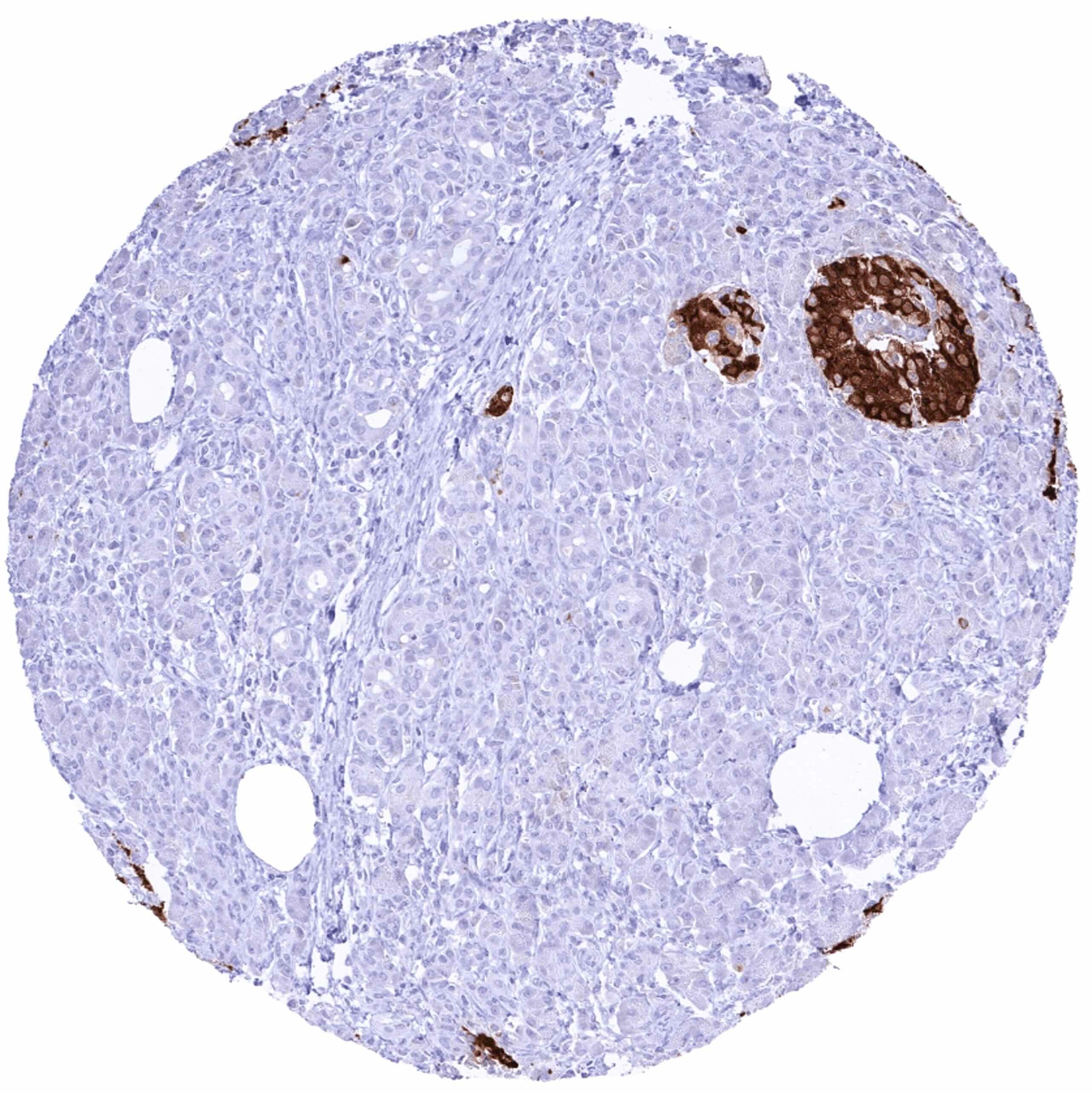
Staining Pattern in Normal Tissues
Manual protocol
Freshly cut sections should be used (less than 10 days between cutting and staining). Heat-induced antigen retrieval for 5 minutes in an autoclave at 121°C in pH 7,8 Target Retrieval Solution buffer. Apply HMV363 at a dilution of 1:150 at 37°C for 60 minutes. Visualization of bound antibody by the EnVision Kit (Dako, Agilent) according to the manufacturer’s directions.
| Brain | Cerebrum | Negative | |
| Cerebellum | Negative | ||
| Endocrine tissues | Thyroid | Negative | |
| Parathyroid | Negative | ||
| Adrenal gland | Negative | ||
| Pituitary gland | Negative | ||
| Respiratory system | Respiratory epithelium | Negative | |
| Lung | Negative | ||
| Gastrointestinal tract | Salivary glands | Negative | |
| Esophagus | Negative | ||
| Stomach | Negative | ||
| Duodenum | Negative | ||
| Small intestine | Negative | ||
| Appendix | Negative | ||
| Colon | Negative | ||
| Rectum | Negative | ||
| Liver, Gallbladder, Pancreas | Liver | Negative | |
| Gallbladder | Negative | ||
| Pancreas | Strong cytoplasmic C-peptide immunostaining of the majority of islet cells. A faint staining of acinar cells surrounding pancreatic islets can be seen because of „contamination artifacts“. | ||
| Genitourinary | Kidney | Negative | |
| Urothelium | Negative | ||
| Male tissues | Prostate | Negative | |
| Seminal Vesicles | Negative | ||
| Testis | Negative | ||
| Epididymis | Negative | ||
| Female tissues | Breast | Negative | |
| Uterus, myometrium | Negative | ||
| Uterus, ectocervix | Negative | ||
| Uterus, endocervix | Negative | ||
| Uterus, endometrium | Negative | ||
| Fallopian tube | Negative | ||
| Ovary | Negative | ||
| Placenta early | Negative | ||
| Placenta mature | Negative | ||
| Amnion | Negative | ||
| Chorion | Negative | ||
| Skin | Epidermis | Negative | |
| Sebaceous glands | Negative | ||
| Muscle, connective & soft tissues | Heart muscle | Negative | |
| Skeletal muscle | Negative | ||
| Smooth muscle | Negative | ||
| Vessel walls | Negative | ||
| Fat | Negative | ||
| Stroma | Negative | ||
| Endothelium | Negative | ||
| Bone marrow & lymphoid tissues | Bone marrow | Negative | |
| Lymph node | Negative | ||
| Spleen | Negative | ||
| Thymus | Negative | ||
| Tonsil | Negative |
Details
More product details
Biology behind
C-peptide (connecting peptide) is a part of the proinsulin protein which is coded by the insulin gene at 11p15.5 and produced exclusively by the beta cells of the pancreatic islets. C-peptide is formed when the proinsulin is split into insulin and C-peptide. At that time equimolar quantities of insulin and C-peptide are released to the blood. C-peptide binds to the surface of several cell types (neuronal, endothelial, fibroblast and renal tubular) and can activate specific pathways. The clinical significance of C-peptide lies in its serological measurement as a parameter for endogenous insulin production (not influenced by exogenous insulin).
Protocol Recommendations
Potential Research Applications
Evidence For Specificity In I H C


