
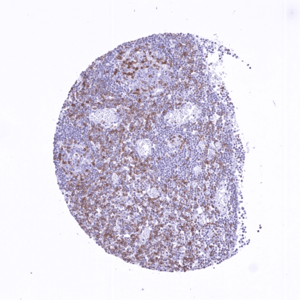
Tonsil showing many TIGIT positive lymphocytes are in the interfollicular area while the strongest TIGIT staining occurs in a subset of lymphocytes in the germinal centre
Staining Pattern in Normal Tissues
Manual protocol
Freshly cut sections should be used (less than 10 days between cutting and staining). Heat-induced antigen retrieval for 5 minutes in an autoclave at 121°C in pH 7,8 Target Retrieval Solution buffer. Apply HMV322 at a dilution of 1:150 at 37°C for 60 minutes. Visualization of bound antibody by the EnVision Kit (Dako, Agilent) according to the manufacturer’s directions.
| Brain | Cerebrum, grey | Negative | |
| Cerebrum, white | Negative | ||
| Cerebellum, cortex | Negative | ||
| Cerebellum, white | Weak to moderate, nuclear staining of a subset of (probaly glial) cells (crossreactivity). | ||
| Ganglion | Negative | ||
| Ependyma | Negative | ||
| Eye, retina | Negative | ||
| Endocrine Tissues | Thyroid | Negative | |
| Parathyroid gland | Negative | ||
| Adrenal gland | Negative | ||
| Pituitary gland, anterior lobe | Negative | ||
| Pituitary gland, posterior lobe | Negative | ||
| Respiratory system | Lung bronchi | Negative | |
| Lung, bronchial glands | Negative | ||
| Nose, paranasal sinus | Negative | ||
| Lung, parenchyma | Negative | ||
| Proximal digestive tract | Lip | Negative | |
| Oral cavity | Negative | ||
| Tonsil, surface | Negative | ||
| Esophagus, mucosa | Negative | ||
| Lip, small salivary gland | Negative | ||
| Sublingual gland | Negative | ||
| Parotid gland | Negative | ||
| Submandibullary gland | Negative | ||
| Gastronintestinal tract | Stomach, antrum | Negative | |
| Stomach, fundus and corpus | Negative | ||
| Small intestine, duodenum | Negative | ||
| Duodenum, Brunner gland | Negative | ||
| Small intestine, ileum | Negative | ||
| Appendix | Negative | ||
| Colon descendens | Negative | ||
| Rectum | Negative | ||
| Anal canal, transition epithelium | Negative | ||
| Liver, Gallbladder, Pancreas | Liver | Negative | |
| Gallbladder | Negative | ||
| Pancreas | Negative | ||
| Kidney, urinary bladder | Kidney, cortex | Negative | |
| Kidney, medulla | Negative | ||
| Urinary bladder, urothelium | Negative | ||
| Kidney pelvis, mucosa | Negative | ||
| Male tissues | Prostate | Negative | |
| Seminal vesicle | Negative | ||
| Epididymis caput | Negative | ||
| Epididymis cauda | Negative | ||
| Testis | Negative | ||
| Female Tissues | Breast, glands | Negative | |
| Ectocervix | Negative | ||
| Endocervix | Negative | ||
| Endometrium, proliferation | Negative | ||
| Endometrium, secretion | Negative | ||
| Uterus, myometrium | Negative | ||
| Fallopian tube | Negative | ||
| Ovary, stroma | Negative | ||
| Ovary, follicular cyst | Negative | ||
| Ovary, corpus luteum | Negative | ||
| Amnion | Negative | ||
| Chorion | Negative | ||
| Amnion/Chorion | Negative | ||
| Placenta, early, decidua | Negative | ||
| Placenta, first trimenon | Negative | ||
| Placenta, mature | Negative | ||
| Muscle, connective & soft tissue | Aorta, intima | Negative | |
| Skeletal muscle | Negative | ||
| Aorta, media | Negative | ||
| Skeletal muscle, tongue | Negative | ||
| Heart, left ventricle | Negative | ||
| Kidney pelvis, muscular wall | Negative | ||
| Urinary bladder, muscular wall | Negative | ||
| Esophagus, muscular wall | Negative | ||
| Stomach, muscular wall | Negative | ||
| Ileum, muscular wall | Negative | ||
| Appendix, muscular wall | Negative | ||
| Colon descendens, muscular wall | Negative | ||
| Penis, glans, corpus spongiosum | Negative | ||
| Fat, white | Negative | ||
| Skin | Skin, surface | Negative | |
| Skin (hairs, sebaceous glands) | Negative | ||
| Anal canal, skin | Negative | ||
| Scrotum | Negative | ||
| Bone Marrow & lymphoid tissues | Bone marrow | Negative | |
| Thymus | Weak to moderate TIGIT staining of a subset of lymphocytes. | ||
| Spleen | Weak to moderate TIGIT staining of a subset of lymphocytes. | ||
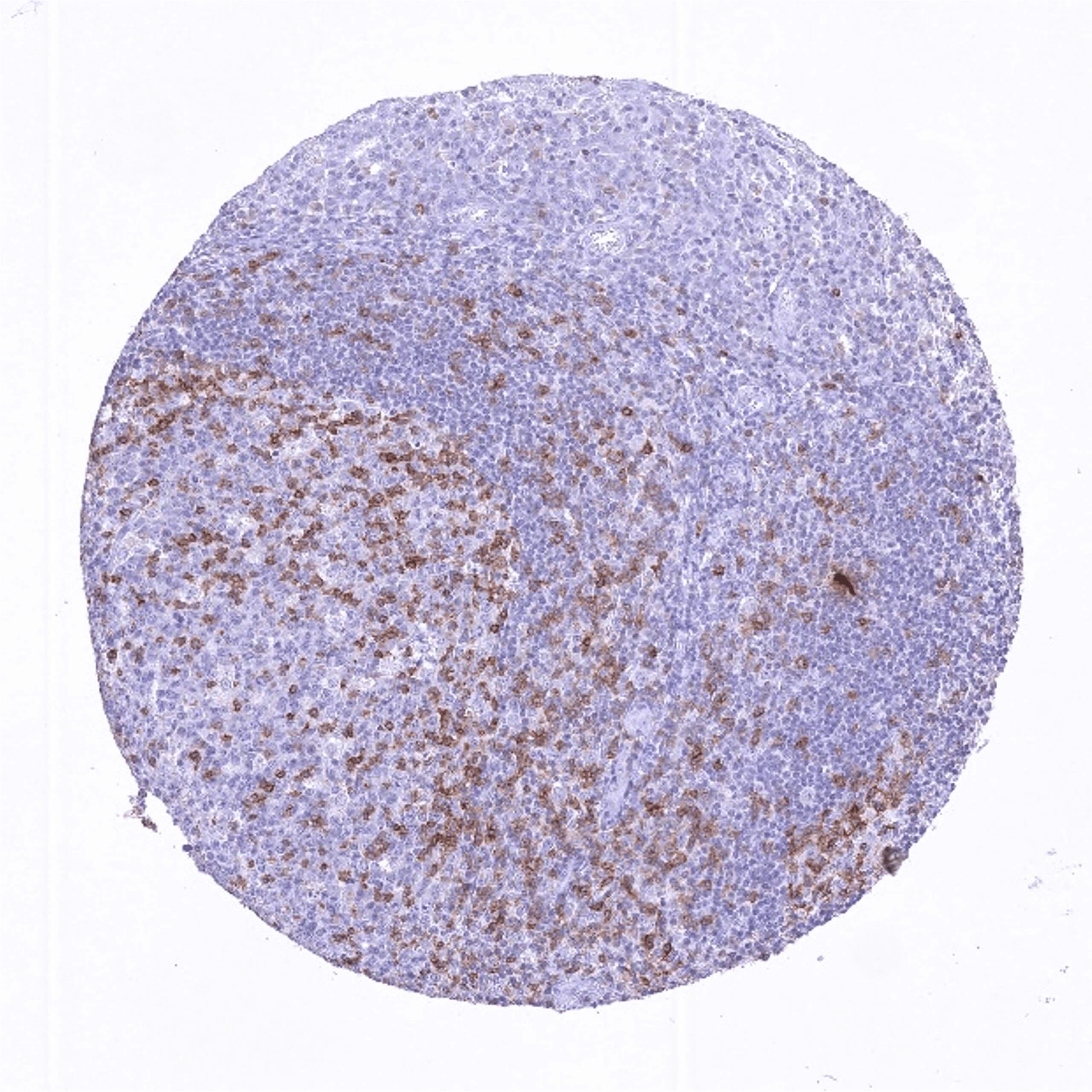
| Lymph node | Most TIGIT positive lymphocytes are in the interfollicular area while the strongest TIGIT staining occurs in the few labeled lymphocytes in germinal centres. | ||
| Tonsil, deep | Variable levels of TIGIT staining in subsets of follicular and interfollicular lymphocytes. The highest level of expression occurs in CD4+ follicular T helper cells located in the germinal centre periphery orientated towards the tonsil surface epithelium. |
Details
More product details
More product details
Biology behind
TIGIT (T cell immunoreceptor with Ig and ITIM domains) is a transmembrane glycoprotein of the poliovirus receptor (PVR) family which is coded by the TIGIT gene at 3q13.31. TIGIT acts as an inhibitory immune receptor (immune checkpoint) which can bind to CD155 with high affinity, and to CD112 with lower affinity. TIGIT expression is restricted to some CD8+ cytotoxic T cells, CD4+ T helper cells, FOXP3+ regulatory T cells, and NK cells. The highest level of expression occurs in CD4+ follicular T helper cells located in the germinal centre periphery orientated towards the tonsil surface epithelium. TIGIT expression has a limiting effect on antitumoral immune reactions. TIGIT inhibition, by either genetic ablation or blocking antibodies, increases T-cell activation and proliferation in response to stimulation and consequently results in reduced tumor growth in experimental models. Various compounds targeting TIGIT have been developed (i.e. tiragolumab, domvanalimab, vibostolimab, etigilimab, m6223, ociperlimab) and are currently evaluated in clinical trials, mostly in combination with other immune checkpoint inhibitors.
Protocol Recommendations
Protocol Recommendations
Potential Research Applications
Potential Research Applications
Evidence For Antibody Specificity In I H C
Evidence For Antibody Specificity In I H C


