Product image 1

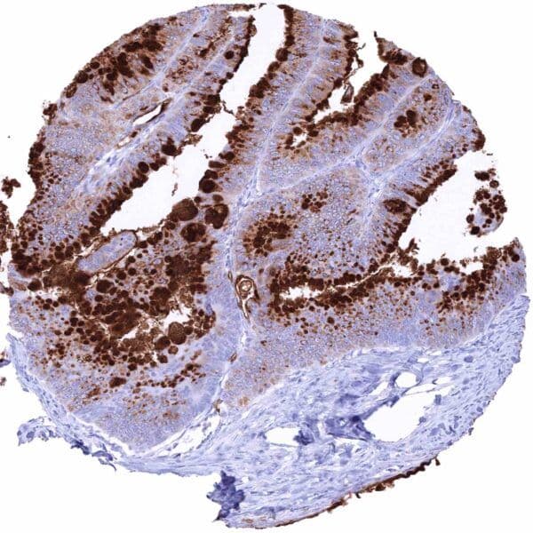
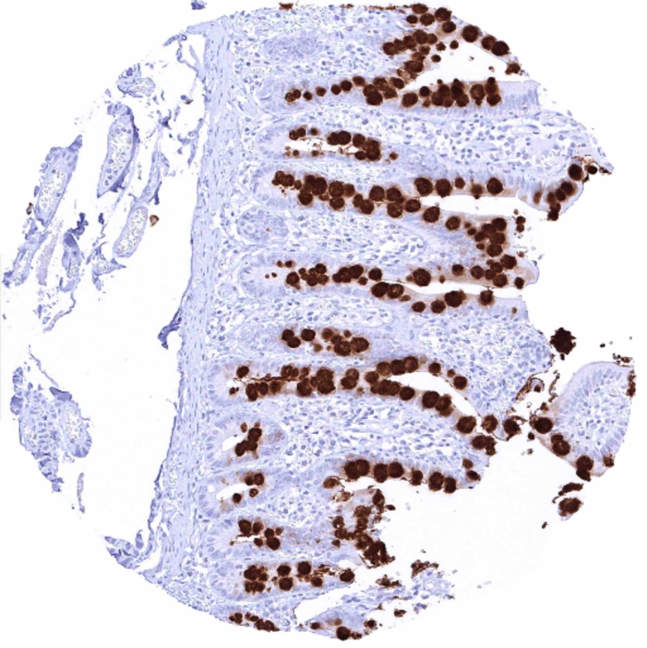
Ileum sample with strong MUC2 positivity of mucins in goblet cells.

Staining Pattern in Normal Tissues

Manual protocol

Freshly cut sections should be used (less than 10 days between cutting and staining). Heat-induced antigen retrieval for 5 minutes in an autoclave at 121°C in pH 7,8 Target Retrieval Solution buffer. Apply HMV310 at a dilution of 1:150 at 37°C for 60 minutes. Visualization of bound antibody by the EnVision Kit (Dako, Agilent) according to the manufacturer’s directions.

BrainCerebrum, grey Negative
Cerebrum, white Negative
Cerebellum, cortex Negative
Cerebellum, white Negative
Ganglion Negative
Ependyma Negative
Eye, retina Negative
Endocrine TissuesThyroid Negative
Parathyroid gland Negative
Adrenal gland Negative
Pituitary gland, anterior lobe Negative
Pituitary gland, posterior lobe Negative
Respiratory systemLung bronchi Negative
Lung, bronchial glands Negative
Nose, paranasal sinus Negative
Lung, parenchyma Negative
Proximal digestive tractLip Negative
Oral cavity Negative
Tonsil, surface Negative
Esophagus, mucosa Negative
Lip, small salivary gland Negative
Sublingual gland Negative
Parotid gland Negative
Submandibullary gland Negative
Gastronintestinal tractStomach, antrum Negative
Stomach, fundus and corpus Negative
Small intestine, duodenum Negative
Duodenum, Brunner gland Strong MUC2 positivity of mucins in goblet cells.
Small intestine, ileum Negative
Appendix Negative
Colon descendens Strong MUC2 positivity of mucins in goblet cells.
Rectum Negative
Anal canal, transition epithelium Negative
Liver, Gallbladder, PancreasLiver Negative
Gallbladder Negative
Pancreas Negative
Kidney, urinary bladderKidney, cortex Negative
Kidney, medulla Negative
Urinary bladder, urothelium Negative
Kidney pelvis, mucosa Negative
Male tissuesProstate Negative
Seminal vesicle Negative
Epididymis caput Negative
Epididymis cauda Negative
Testis Negative
Female TissuesBreast, glands Negative
Ectocervix Negative
Endocervix Negative
Endometrium, proliferation Negative
Endometrium, secretion Negative
Uterus, myometrium Negative
Fallopian tube Negative
Ovary, stroma Negative
Ovary, follicular cyst Negative
Ovary, corpus luteum Negative
Amnion Negative
Chorion Negative
Amnion/Chorion Negative
Placenta, early, decidua Negative
Placenta, first trimenon Negative
Placenta, mature Negative
Muscle, connective & soft tissueAorta, intima Negative
Skeletal muscle Negative
Aorta, media Negative
Skeletal muscle, tongue Negative
Heart, left ventricle Negative
Kidney pelvis, muscular wall Negative
Urinary bladder, muscular wall Negative
Esophagus, muscular wall Negative
Stomach, muscular wall Negative
Ileum, muscular wall Negative
Appendix, muscular wall Negative
Colon descendens, muscular wall Negative
Penis, glans, corpus spongiosum Negative
Fat, white Negative
SkinSkin, surface Negative
Skin (hairs, sebaceous glands) Negative
Anal canal, skin Negative
Scrotum Negative
Bone Marrow & lymphoid tissuesBone marrow Negative
Thymus Negative
Spleen Negative
Lymph node Negative
Tonsil, deep Negative

MUC2

(HMV310)

MUC2 is a main protein of intestinal mucus barrier.

Select Format
Select Volume
From €295.00
excl. shipping costs and taxes
MUC2 (HMV310)
€295.00

Details

Type
Recombinant Rabbit monoclonal / IgG
Clone
HMV310
Reactivity
Human

More product details

Biology behind

Mucin 2 (MUC2) is an oligomeric mucus gel-forming protein coded by the MUC2 gene at chromosome 11p15.5. The protein is secreted by intestinal goblet cells to the gut where it constitutes the main component of colorectal mucus. The mucins function as a defense mechanism to maintain the integrity of the epithelial cells which are continuously exposed to luminal contents that include large quantities of different bacteria, proteases, bile, and ingested toxins. The mucin barrier consists of two layers. The inner layer is directly attached to the epithelium, densely packed, largely consists of uncleaved MUC2, and is free from bacterial colonization. The outer layer contains bacteria and is less dense due of proteolytic cleavage of MUC2. Under normal conditions, the protease-resistant mucus layer prevents intestinal bacteria to make physical contact with epithelial cells by virtue of its only minute pore sizes and strong hydrophobic properties capable to repel bacteria in the aqueous lumen. Loss of MUC2 confers a microenvironment in which bacteria can activate an inflammatory response at the epithelial surface. The defensive function of MUC2 and of other mucins to maintain the integrity of the epithelial cells can also be exploited by tumor cells in limiting the activation of inflammatory responses.

Protocol Recommendations

Potential Research Applications

Evidence For Specificity In I H C